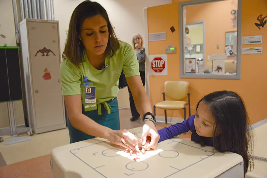

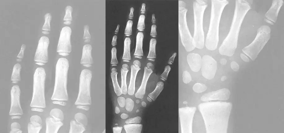
When doctors want to understand how a child is growing and developing, they sometimes order a bone age exam. This simple X-ray of the hand and wrist helps doctors estimate how mature a child’s bones are compared with other children of the same age.
A new study published in Pediatric Radiology evaluated three advanced artificial intelligence (AI) tools that estimate bone age. Authored by Dr. Elanchezhian Somasundaram, Dr. Rama Ayyala, William Tepe, Bryan Luna, and Dr. Jonathan Dillman, the findings could help improve the accuracy and fairness of bone age assessments for children in the future.
A bone age exam is a low-dose X-ray, usually of the left hand and wrist. Doctors use it to help evaluate:
The bone age result is compared with a child’s actual age (called chronological age) to help determine whether growth and development are occurring as expected.
Think of bone age as a way to see a child’s “growth clock.” Two children may both be 12 years old, but one child’s bones may look more like those of an 11-year-old while another’s may resemble those of a 13-year-old. This information can help doctors understand growth patterns and make important treatment decisions.
Researchers from Cincinnati Children’s Hospital Medical Center evaluated more than 7,100 children between the ages of 2 and 18 years.
Instead of looking at children with growth disorders, they studied children who had normal hand X-rays after injuries. This allowed researchers to examine bone age estimates in a large group of contemporary, generally healthy children.
The team tested three leading AI systems designed to estimate bone age and compared the AI predictions with each child’s actual age.
The study found that all three AI systems consistently estimated children to be slightly older than their actual age.
On average:
While these differences may seem small, they were consistent across thousands of children.
Many bone age assessments are based on standards created using children’s X-rays collected in the 1930s and 1940s. Researchers believe that modern children may mature somewhat differently than children did nearly a century ago. Factors such as nutrition, healthcare, environment, and overall health may contribute to these differences. This study suggests that some of the standards used today may not fully reflect the growth patterns of children growing up in the 21st century.
Growth-related decisions often depend on bone age results. More accurate assessments can help doctors better evaluate growth concerns, make more informed treatment recommendations, improve timing of medical interventions, and reduce the chance of unnecessary worry or testing. Understanding where current methods may have limitations is an important step toward improving care.
Artificial intelligence has the potential to make bone age assessments faster, more consistent, and more accessible. However, this study highlights an important lesson: AI tools must be tested on diverse, real-world populations before being widely adopted. Even highly sophisticated AI systems can reflect limitations in the historical data they were trained on.
One particularly important finding was that the amount of overestimation varied among different groups of children. The researchers observed differences related to sex, race, ethnicity, and age. This is important because every child deserves accurate and equitable medical care. Studies like this help researchers identify where improvements are needed so future tools work well for all children, regardless of background.
Not at all. Bone age remains a valuable and widely used tool in pediatric medicine. The differences found in this study were generally modest, and bone age is only one piece of the overall clinical picture.
Doctors never make decisions based on a bone age result alone. They also consider your child’s height and weight, growth history, physical examination, family growth patterns, and laboratory results when needed. The findings simply help researchers and clinicians understand how to make these tools even better.
This study is good news for patients and families because it shows researchers are actively working to improve how children’s growth is assessed.
The findings may lead to:
Bone age exams are an important part of evaluating children’s growth and development. This large study found that current AI-based bone age tools often estimate children as slightly older than their actual age, suggesting that modern children may not perfectly match the historical standards used for decades.
For families, the key takeaway is positive: Researchers are identifying ways to make growth assessments more accurate, more equitable, and better tailored to today’s children. Ultimately, this work supports the goal every parent wants—providing the best possible care for every child.

Glenn Miñano is a media specialist in the Department of Radiology, providing graphic design, photography, printing, video services, and administration of the department’s online properties. His works have been published in several medical articles, such as the American Journal of Radiology and the American Institute of Ultrasound. He has been providing these services to the Radiology Department since 1996.

Editor: Meredith Towbin