
Based on research by Mishka G. Hoo Kim, MBBS, Cara E. Morin, MD, PhD, Susan E. Sharp, MD, and Andrew T. Trout, MD, published in Academic Radiology.
For children living with hydrocephalus or other conditions that require a cerebrospinal fluid (CSF) shunt, concerns about shunt malfunction can be stressful for both patients and families. When symptoms such as headaches, vomiting, irritability, sleepiness, or changes in behavior occur, doctors may need to determine whether a shunt is working properly. One of the tools they use is a nuclear medicine CSF shunt study, which helps evaluate how fluid moves through the shunt system.
A new study from Cincinnati Children’s Hospital examined five years of shunt imaging exams and found something important: Most children receive the information doctors need within the first 60 minutes of imaging. This finding could help reduce testing times, improve efficiency, and get families answers sooner.
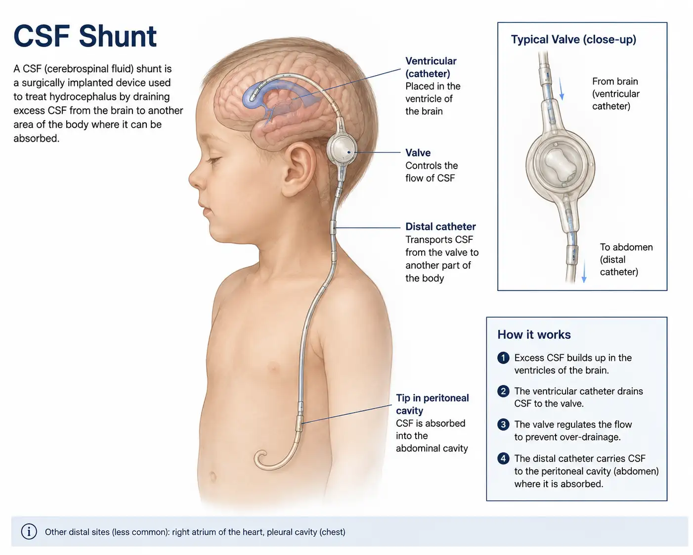
A CSF shunt is a small medical device that helps drain excess cerebrospinal fluid from around the brain or spinal cord. The shunt redirects fluid to another area of the body where it can be absorbed safely, such as the abdomen (ventriculoperitoneal or VP shunt), the chest cavity (ventriculopleural or VPL shunt), or the bloodstream (ventriculoatrial or VA shunt). Shunts can be lifesaving, but they can occasionally develop problems such as blockages or mechanical failures.
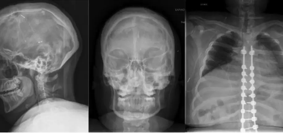
When doctors suspect a shunt problem, they often start with imaging such as MRI, CT scans, ultrasound, and an X-ray shunt series. Sometimes those tests don’t provide a clear answer.
A nuclear medicine CSF shunt study offers something different: It shows how well fluid is actually moving through the shunt, helping doctors assess its function. During the exam, a tiny amount of radiopharmaceutical is placed into the shunt system, and special images track its movement through the tubing. If the tracer reaches the expected drainage location, it suggests that portion of the shunt is working.
The researchers reviewed:
The study included infants, children, and young adults. The goal was to determine how long it typically takes for the tracer to travel through the shunt and reach its destination. Understanding this timing could help optimize future imaging protocols.
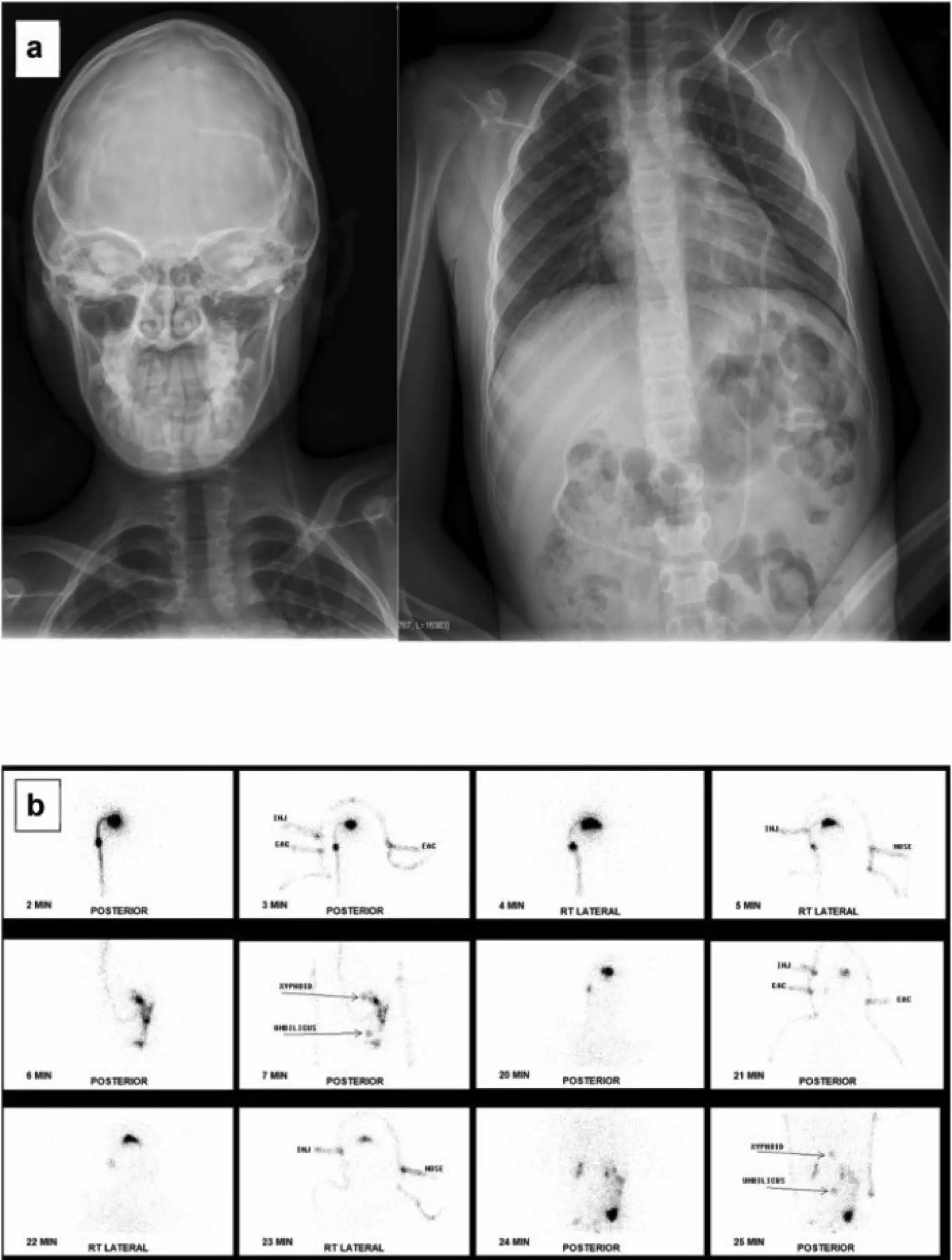
The good news is that in the vast majority of cases, doctors were able to see the tracer reach the drainage area within 60 minutes.
Ventriculoperitoneal (VP) shunts were the most common type studied. Researchers found the median time to reach the abdomen was 17 minutes. Most showed activity far earlier than 60 minutes, and only 6 out of 174 VP shunts showed activity after 60 minutes. Ventriculopleural (VPL) shunts drain fluid into the chest cavity. Researchers found the median time to reach the abdomen was 5 minutes. Ventriculoatrial (VA) shunts drain into the bloodstream. The median time to reach the abdomen was 7.5 minutes.
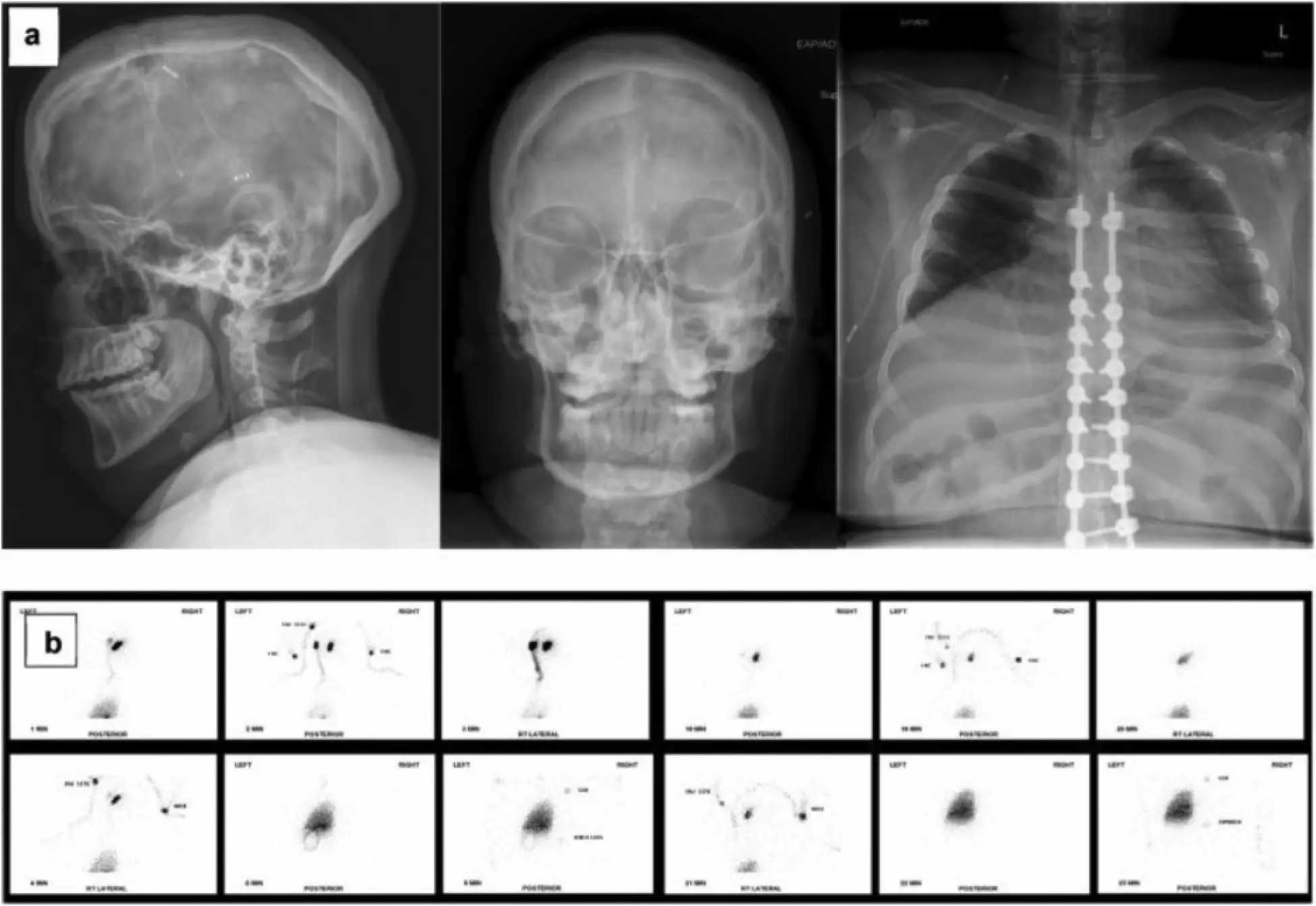
In most patients, valuable information became available relatively quickly.
One of the biggest benefits of this research is the possibility of reducing how long some children need to remain in the imaging department. If future protocols confirm these findings, many patients may not need extended imaging sessions beyond 60 minutes. This could mean less waiting, more comfort, more convenient appointments, and reduced disruption to school and family schedules
This research suggests that doctors can often obtain the information they need earlier in the examination, paving the way for faster clinical decisions. Faster imaging results can help support faster diagnosis and treatment planning, more timely neurosurgical decisions, and reduced uncertainty for families.
Although nuclear medicine shunt studies are generally well tolerated, shorter imaging times can also help reduce anxiety, discomfort, and boredom during testing. Any opportunity to make testing easier is a win for pediatric patients.
The study helps healthcare teams better understand what “normal” tracer movement looks like, creating more efficient care. By knowing when activity typically appears in different shunt types, radiologists can recognize abnormalities more confidently, optimize imaging schedules, improve department efficiency, and focus resources where they are most needed. Ultimately, this helps improve the patient experience.
Not necessarily. An important finding from the study was that some children still required surgery even when the tracer reached the drainage area. Why? Because doctors consider many factors, including symptoms, physical examination, MRI or CT findings, X-rays, shunt function studies, and neurosurgical evaluation. A shunt study is one important piece of the puzzle, but it is not the only factor used in decision-making.
This study provides reassuring information for families whose children undergo nuclear medicine shunt imaging. Researchers found that:
? Most shunts that are functioning show expected fluid movement within 60 minutes.
? Longer imaging sessions rarely provided additional information.
? Understanding shunt flow timing may help improve future imaging protocols.
? More efficient testing could reduce waiting, anxiety, and appointment length.
? Faster results may help support quicker treatment decisions.
For children and young adults with CSF shunts, nuclear medicine imaging plays an important role in evaluating whether a shunt is working properly. This five-year study found that the vast majority of useful information is obtained within the first hour of imaging.
For families, that means a future with potentially shorter exams, faster answers, and a more streamlined care experience. While every child is unique and imaging decisions remain individualized, this research is an important step toward making shunt evaluations more efficient, comfortable, and family-friendly.

Glenn Miñano is a media specialist in the Department of Radiology, providing graphic design, photography, printing, video services, and administration of the department’s online properties. His works have been published in several medical articles, such as the American Journal of Radiology and the American Institute of Ultrasound. He has been providing these services to the Radiology Department since 1996.

Editor: Meredith Towbin