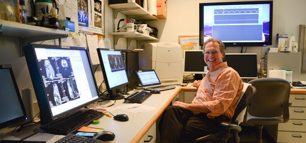

I started my medical imaging career in 1991, as an x-ray technologist working in the Emergency Room from 4 pm to midnight. Soon, I was asked to start working with this new way of taking pictures without x-rays, which was magnetic resonance imaging, or MRI. Five years later, I was a charter member of the new Imaging Research Center (IRC) at Cincinnati Children’s. My MRI education continued for another 24 years while I was part of a team developing new MRI methods that provided diagnostic information without exposing anyone to x-rays.
These new methods measured the extent of juvenile rheumatoid arthritis in children’s joints, how much damage was caused by a childhood stroke, what parts of the young brain were involved in language development, and whether we needed to treat a child for an infection or a tumor. Some of these methods took five years to develop, but MRI research has made a big difference in the outcome for many of our children.
One of the best examples I can think of in which MRI research made a difference is how children were tested to diagnose a blockage in the brain. Back in 1973, this test took most of the day, was very painful, and had bad side effects. A CT scan would replace this test, but there was still a lot of x-ray exposure. With MRI, the blockage can be seen after a 3-minute scan. Your child need only lie still for as long as a television commercial takes and there is no x-ray.
Today, I don’t have the pleasure of working with children. Instead, I work on a very powerful, pre-clinical MRI. We hope to ask the questions that MRI will help us to find the answers to five to ten years from now. I’ll read about it in magazines when I’ve come home from riding bicycles with my grandkids or turning over creek rocks with them just to see what might be underneath.