A recent study led by Dr. Elanchezhian Somasundaram, PhD, from Cincinnati Children’s Radiology Department, has been featured in the American Journal of Roentgenology. The study focuses on using deep learning to improve the accuracy of organ segmentation in pediatric CT scans. By training and validating AI models on both internal hospital data and diverse public datasets, the research demonstrates that transfer learning techniques outperform existing segmentation methods, particularly for more challenging organs like the pancreas. These advancements could lead to better volumetric assessments and more precise treatment planning for children.
Background
Deep learning, a type of artificial intelligence (AI), has been highly effective in detecting and analyzing abdominal organs in adult CT scans. However, research in pediatric cases has been limited. Unlike adults, children’s organs vary significantly in size, shape, and anatomical positioning at different developmental stages, making it challenging for existing AI models to generalize accurately. This study aimed to develop and test deep learning models specifically for children, focusing on the liver, spleen, and pancreas.
Study Objectives
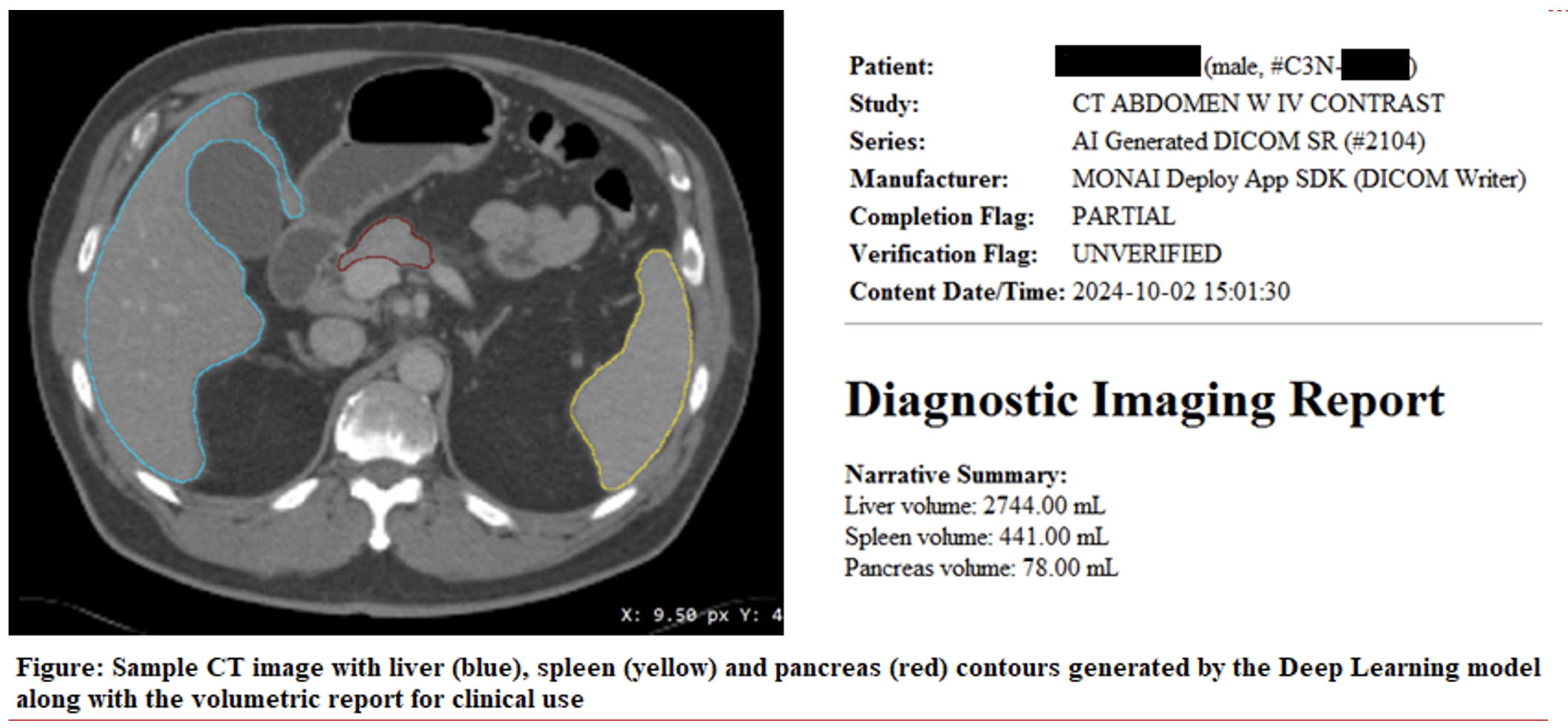
The primary goal of this research was to create and validate AI models that could accurately identify and segment the liver, spleen, and pancreas in pediatric CT scans. By enhancing segmentation precision, these models could assist radiologists in analyzing scans more efficiently, leading to faster diagnoses and improved treatment planning for children.
Methods
The research team trained and tested their AI models using 1,731 CT scans from different sources. These scans included:
Three deep learning models—SegResNet, DynUNet, and SwinUNETR—were trained using two different methods:
The study compared these models with TotalSegmentator, a publicly available AI segmentation tool, to see which performed best.
Results
The AI models were tested on both internal (pediatric) and external (public) datasets. The key findings included:
The best-performing model, DynUNet TL, was chosen for public use and is now available as an open-source tool for further research in MONAI’s model zoo.
Clinical Impact
This research is a major step forward in pediatric imaging. Accurate AI models can help radiologists analyze CT scans more efficiently, leading to better diagnosis and treatment for children. The study’s findings suggest that deep learning can be a valuable tool for pediatric radiology, especially when models are trained with diverse datasets. Currently, this model is being tested for clinical deployment to enhance segmentation workflows for children that undergo abdominal CT.
Dr. Somasundaram and his team’s work highlights the importance of AI in medical imaging and demonstrates Cincinnati Children’s commitment to innovation in pediatric healthcare.
Glenn Miñano is a media specialist in the Department of Radiology, providing graphic design, photography, printing, video services, and administration of the department’s online properties. His works have been published in several medical articles, such as the American Journal of Radiology and the American Institute of Ultrasound. He has been providing these services to the Radiology Department since 1996.
Editor: Meredith Towbin
Elanchezhian Somasundaram, PhD, is a dedicated researcher specializing in translational artificial intelligence (AI) for pediatric healthcare. With a background in computational physics, statistics, and engineering, combined with expertise in pediatric imaging, Elanchezhian develops AI-driven quantitative solutions to enhance clinical diagnostics and improve the quality of care for children.