
What is it?
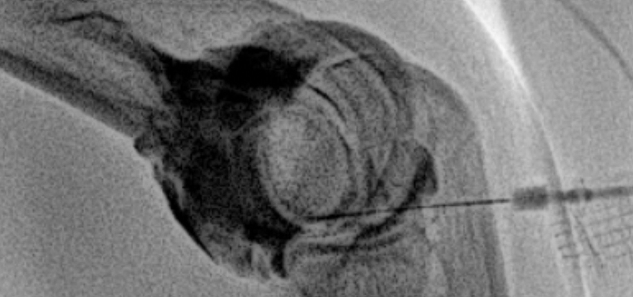
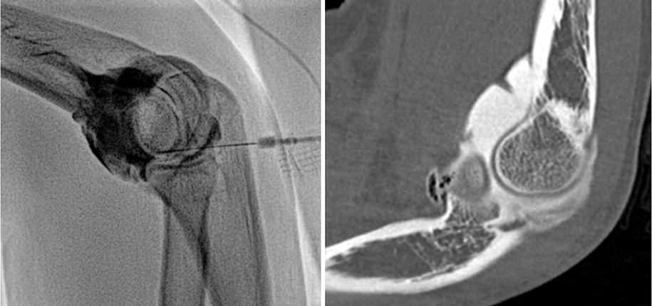
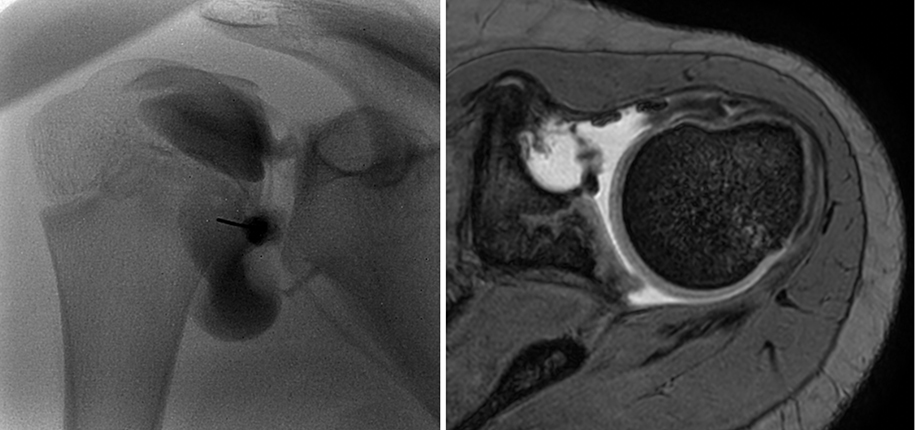
An MR or CT arthrogram is a special test your doctor may order to better evaluate the cause of joint symptoms. Contrast material is injected into a joint (hip, shoulder, wrist, elbow, etc.) The joint is then evaluated with MRI or CT.
Why do it?
The contrast material allows for distension of the joint to enhance the imaging of its internal structures. This allows better visualization of the ligaments and cartilage of the joint to aid the radiologist in making a more accurate diagnosis regarding the cause of the symptoms.
How is it done?
The arthrogram is performed in Interventional Radiology by an interventional radiologist. The MRI or CT is then interpreted by a specially trained musculoskeletal radiologist.
The patient is first brought to interventional radiology. After he or she is appropriately positioned on the x-ray table, the skin is cleaned. Next, the skin is numbed to minimize any discomfort related to the injection. Using x-ray or sometimes an ultrasound, a small needle is then inserted into the joint. Once the needle is in the appropriate position, the contrast material is injected into the joint. After the contrast material is injected, the needle is removed and a bandage is placed over the needle site. The patient is then taken to the MRI or CT scanner where the scan is performed. Following the MRI or CT, the patient is discharged home.


Most of these procedures are performed while patients are awake. Parents are allowed into the room where the injection is performed to help comfort the child. They may watch a movie or listen to music to help distract them during the injection. The interventional radiology staff in the room during the injection are attentive to patients’ needs and will also help comfort them during the injection. In rare cases, patients may require sedation or anesthesia. The Interventional Radiology schedulers and nurses can help determine these needs during the scheduling process.
Contributed by Dr. Manish Patel and edited by Michelle Gramke, Adv Tech-ULT.
