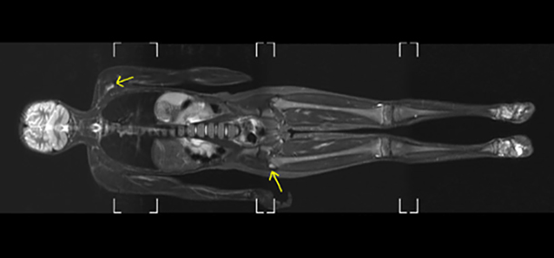

Six-year-old girl with two sites of infection demarcated by the bright appearance, one near the hip and the other near the shoulder (arrows).
One of the many challenges when scanning children with difficult abnormalities or diagnoses are that there are several circumstances in which more than one body part, in the same patient, needs to be evaluated. For this reason, an imaging system capable of performing whole-body magnetic resonance imaging (MRI) can be extremely helpful and a major time saver.
Whole-body MRI can image a child from head to toe without the use of ionizing radiation. This is particularly useful for children who require sedation or general anesthesia in order to undergo the examination. This technique allows an evaluation of the whole body to be performed at one time, in one visit. Your child remains in the same position and information is acquired at different “stations” or “stops” (the table the patient lies on moves), and then these “stations” are reconstructed into a single whole body image. This technique can be utilized regardless of the size of the patient, and has been successful in babies as well as full-sized adults.
The applications for whole-body MRI continue to grow. Currently, the technique is utilized to evaluate both benign and malignant conditions. Some of the uses include the determination of multiple sites of infection or the extent of vascular malformations, the severity of inflammatory disorders, screening for possible tumor in patients with risk factors, and the evaluation of sites of metastatic disease both for initial detection, as well as for the response to treatment. However, when fine detail of a specific abnormality or area, such as a joint, is desired, a dedicated focal examination should be obtained (i.e. knee MRI).
A normal whole-body scan in a full-sized 20-year old man.
Also, there are instances in which a whole-body MRI will be ordered, but we only scan your child from hip to toe or shoulder to fingers. We do still label these exams as whole-body MRIs because multiple joints (hip, knee, and ankle joints or shoulder, elbow, and wrist joints) are being covered.
If there are ever any questions about what is being included in your child’s exam, please feel free to ask your MRI technologist who is taking the pictures and he or she will be more than willing to go over coverage with you. Explaining the procedure and instructing you through the exam are just a few of the roles we fulfill when you come and visit us in Cincinnati Children’s Radiology.
Contributed by Dr. Tal Laor and edited by Tony Dandino (RT, MRI).