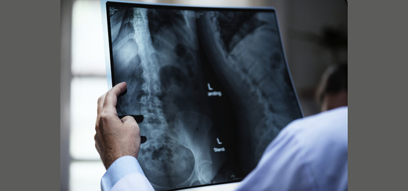
Back when I first started working (over twenty years ago) in the Department of Radiology at Cincinnati Children’s, film was being used to practically capture all radiology modality images. In CT, film is is used to document any head injuries, while in ultrasound it’s used to show a static image of the kidneys or unborn child, among other things. This is by no means a detailed historical study of the rise and fall of black and white film in radiology, but a perspective of one that has seen film being surpassed by technology.
In radiography, x-ray was exposed on large film sheets in order for the radiologist to diagnose a body part. When exposed correctly, film gave a detailed image of the body or foreign objects inside of the body. The technologist would expose the blank film contained in a flat container, called a “cassette” and take that cassette to a darkroom to process the film. Every day hundreds of black and white films were being exposed and developed. The department had more than one dark room to process the films.
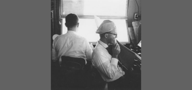
 Photo: Pre 1990’s, historical image of an x-ray View Box (background). Future machines would be larger and automated.
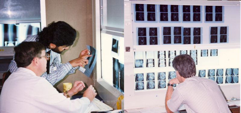
The radiologists would read these film studies on a large vertical lightbox-type machine. The machine would be about five feet wide and as tall as the room. File Room staff would come in and load the machines with the daily x-ray films for the radiologists to ready.
 Photo: Radiologists reading film on the large View Box machines.
There are downsides to everything and film was not an exception. Film contained silver, a precious metal, which made it expensive. The chemicals used to process the film were also expensive and bad for the environment.
Film took up a lot of space in records keeping, hence the need for a file room. These films would be kept in a large envelope call “jackets.” File Room staff would be responsible for organizing and keeping track of the film jackets.
There was only one original, unless the film was copied; once the original was gone, it was gone. It would take a lot of man hours to look for a lost film study, due to mishandling.
It was heavy. Normally, more than one x-ray exposure was taken of a body part to insure the radiologist can diagnose the study at different angles. Over a patient’s lifetime health record, their x-ray jacket would weigh over a pound or so.
As medical technology became better and better, I started to see less use of film in radiology modalities. Once digital scanners were able to capture highly detailed medical images and monitors were able to present high-quality images that rivaled film and software (such as PACS) that gave radiologists the ability to quickly view, diagnose and organize studies, it was only a matter of time film would be another excerpt in history.
Today in our radiology department, we have moved from film to digital. Our File Room is now our large conference room. Our File Room staff have been re-trained to be our Reading Room assistants. The darkrooms are gone, giving us more space. The large bulky x-ray light boxes that took up so much space in the reading room were replaced with medical grade monitors, giving up to four radiologists to work in one room. I’m interested to see what else evolves in radiology in the future to come.