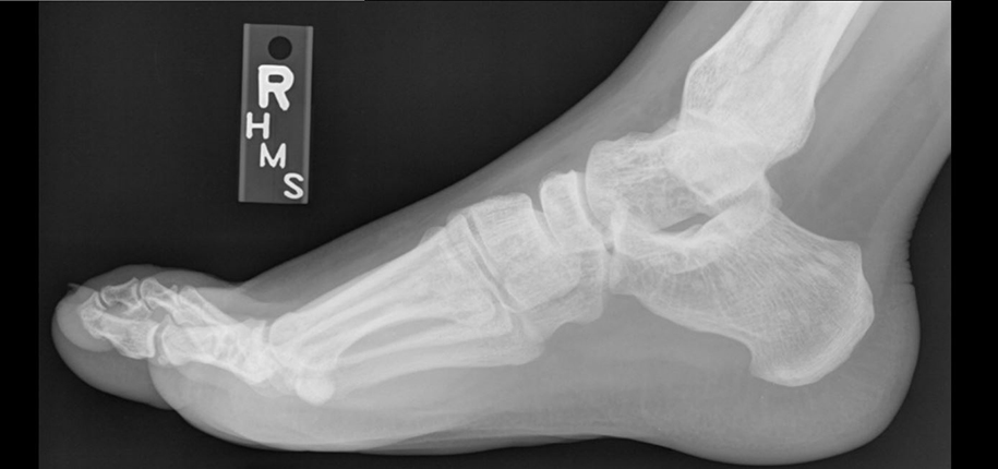

There are several ways that radiologists make images of patients, but the oldest technique and one of the most widely known is the x-ray. The first image made using x-rays was produced in 1895 by Wilhelm Roentgen. But what exactly goes into creating those images?
We’re all used to using visible light to take pictures. In fact, since most cell phones have cameras, there’s a good chance you have a camera with you right now! When you visit our Radiology Department to have an x-ray exam, we use a special kind of camera to take a picture of the part of your body your doctor needs us to look at. To make it less confusing, radiologists call the beam of energy used to take these pictures an x-ray and the picture made using that beam of energy the radiograph.
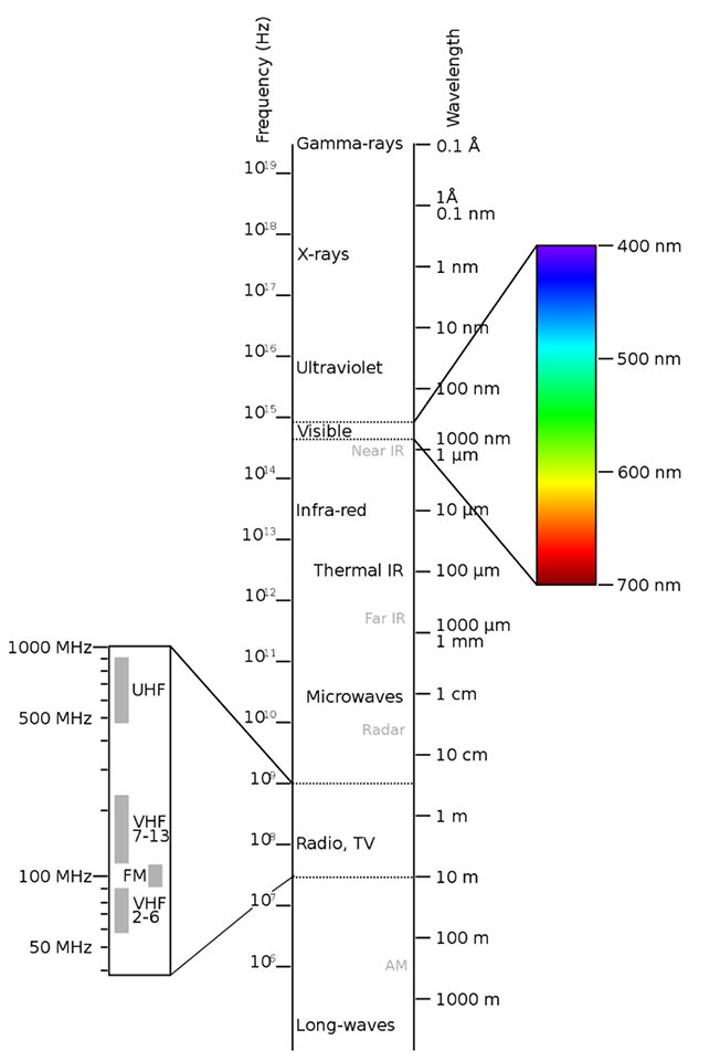
To understand what x-rays are, let’s talk about how they’re similar to light. Just like visible light travels from the sun through space to light up the earth, many other kinds of energy do the exact same thing. All these kinds of energy together make up the electromagnetic spectrum, and radiologists use several of the kinds of energy that we can’t see with our eyes to look inside the human body.

In radiology:
We’re used to the ways visible light interacts with the world around us. Some things are transparent, or see-through, like a window. Other things are translucent, where some of the light passes through and some is blocked, like a lampshade. Other things are opaque and block light, like metal, wood, or a person. Just like visible light, x-rays are blocked more by some materials than by others. In the human body, bones block x-rays very well, while skin and muscle block them less. Parts of the body that contain air, like lungs, block x-rays even less. The amount of blocking, or attenuation, of the x-ray beam is related to density. Generally, the more dense an object, the better it blocks the x-ray beam.
When an x-ray beam passes through a person, we can collect the energy on the other side to see what body parts blocked the beam. Just like the first visible light cameras used film to capture an image, the first x-ray cameras did, too. When the x-ray beam hit the film, it changed the color of the film, and that allowed the x-ray to be turned into a picture that could be seen with the eye. Just like most cameras we use these days are digital, though, so are many modern x-ray systems.
Pictures made using x-rays help doctors make many important diagnoses. They can help us see pneumonia in the lungs or a broken bone, just to name two examples.
Being a radiologist doesn’t give you superpowers, but with a little help from science, we can use x-rays and other techniques to see inside the human body, which seems pretty close!
Dr. Stacey Elangovan, author; Glenn Miñano, editor; Meredith Towbin, copyeditor.