Radiating Hope: Radiology Department Blog

Search

Using X-Rays To Evaluate Growth

Using X-Rays To Evaluate Growth

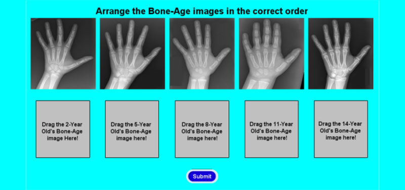
An x-ray of the left hand and wrist is often used to evaluate the skeletal maturity of a child or teenager. The level of skeletal maturity can then be compared to the patient’s chronological age to see if they are growing faster or slower than expected. The x-ray allows doctors to see growth centers which change in number, size, and shape as the patient ages.

Can you put the following bone age x-rays in the correct order from the youngest to the oldest patient?

Contributions by Dr. Susan Sharp and edited by Tim O’Connor, (Informatics Director).

Sharp template

Avatar photo

About the author: Alex Towbin

Alex is a radiologist and the Neil D. Johnson Chair of Radiology Informatics. In this role, he helps to manage the information systems used by the Radiology department. Clinically, Alex is the Assistant Director of thoracoabdominal imaging. His research interests include liver disease, liver tumors, inflammatory bowel disease, and appendicitis.

About The Department

The Radiology Department at Cincinnati Children's is a leader in pediatric diagnostic imaging, radiology research, and radiation dose reduction.

Radiology Specialties