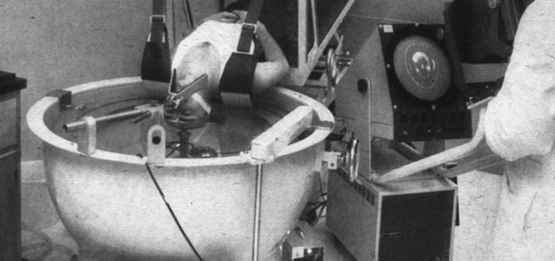
Sounds crazy doesn’t it! But that is how ultrasound imaging started back in the 1950s. The image you see was used to look at the patient’s brain with ultrasound. If the abdomen was being imaged, the patient would have to get more than just their head wet! He or she would be immersed in a water bath, similar to a hot tub, but without any jets of swirling water, and the ultrasound probe would be moved across the abdomen. The technology was so amazing at the time that it was featured in Life Magazine in 1954.
With advances in technology, the imaging probes are much smaller, handheld, and no longer require a water bath to transmit the ultrasound beam. Better ultrasound coupling agents were developed, first using oil and now ultrasound gel. In the 1950s the equipment needed to generate the outgoing ultrasound pulse and to detect the returning ultrasound beam could fill a large closet. Now we have portable machines as small as your smart phone or computer tablet. And the images that are produced by modern ultrasound technology are remarkably detailed, some even producing three-dimensional images in real time, also known as 4D imaging. This type of imaging is very commonly used in obstetrics to monitor babies’ health, especially for twins, triplets, etc.
The best part of ultrasound imaging that remains unchanged over the past 60 years is its safety. Ultrasound does not use x-rays or ionizing radiation to form its images. Radiation exposure should be minimized in all patients, especially young children. Ultrasound is painless and can be safely used on our most fragile patients, including fetuses. And now if your child needs one, he or she doesn’t have to sit in a bath!
Contributed by Dr. Sara O’Hara and edited by Glenn Miñano.