One of the more unique experiences I have had during my year of fellowship was the opportunity to act as one of the ultrasound technologists. I have lots of experience reading ultrasound exams, but not nearly as much when it comes to obtaining the images myself. Think about your favorite movie, one you can quote by heart. Now pretend you’ve been asked to act that movie out. It may be more difficult than you think. I would like to share a bit of information about ultrasound, how it gets used, and how it all affected me.
There are so many applications of ultrasound
As ultrasound doesn’t use radiation, it is often the first test we use to image a patient. It is also one of the few imaging modalities that lets us see the body as it moves. We can watch the blood flow through the vasculature, and the muscles and tendons slide against each other as the patient flexes. It can be very useful, and is often easy for even babies to tolerate. A glob of gel is placed over the area we need to see, and the probe is pressed against the skin, to see inside. The things we can see with ultrasound may prevent most patients from needing exams that come with a radiation cost, like CT.

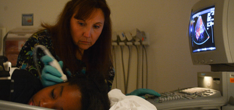
One specific application we use is transcranial Doppler ultrasound, which is used for monitoring in sickle cell patients. These exams are challenging, and often require the patient to cooperate. This mostly boils down to holding their head completely still for 20 minutes or so. I remember the last time my three-year-old held his head still for that long. He was asleep! It is often a group effort to keep an energetic child distracted and calm enough to complete this exam, and I have a new respect for the technologists at Cincinnati Children’s for accomplishing this task every day.
Ultrasound is portable, because it has to be

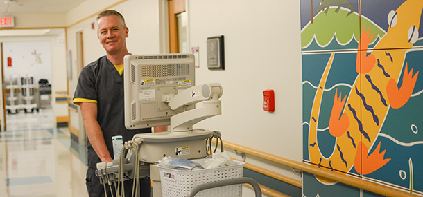
Many patients are too sick to be able to come down to the ultrasound department, so we have to bring ultrasound machine to them. As a temporary ultrasound tech, I was able to assist on intraoperative cases with the kidney transplant surgeons in the OR. The transplant surgeries are very delicate, and it is important for surgeons to see that blood is flowing into and out of the patient’s new kidney or liver as soon as it is “installed.” It is an important collaboration between surgery and radiology, and it can be critical to improving the patient experience.
Beyond the OR, the ultrasound techs and I were also called to the NICU to evaluate the premature infants for head or abdominal ultrasounds. In many cases, these exams are repeated over the course of the hospital stay to monitor for changes and to help the primary care teams provide better patient care.
Ultrasound is also a high priority resource for the emergency room. The indications range from head bumps to foot swelling. In helping out here, I can often help explain to the patients and their parents what I am looking for and what I see. Communication with the family can usually ease some of the anxiety parents are feeling regarding their sick child. It is often helpful for them to ask me questions directly, and I look forward to spending that time with them.
Ultrasound is becoming more commonly used in smaller hospitals and outpatient clinics
On the whole, the experience has been very positive for me. I have been able to spend a lot more time in direct communication with patients and their families, and hopefully made their experience better. I was also able to increase my manual dexterity when scanning by myself. As my year comes to a close, when I look back at what I have learned, I know my experiences here will make me a better pediatric radiologist.
Contributed by Dr. David Hunte and edited by Glenn Miñano, BFA
