When I was in medical school, I remember learning about different illnesses and how to treat them. However, I learned, or better to say, I knew very little compared to what I know now about fetal imaging and things that can be done for babies in need of help to survive in the womb, during the process of birth, or afterward. It seems to me that it was not long ago, but in fact, it has been decades! Many things have changed since then thanks to research and progress in technology.
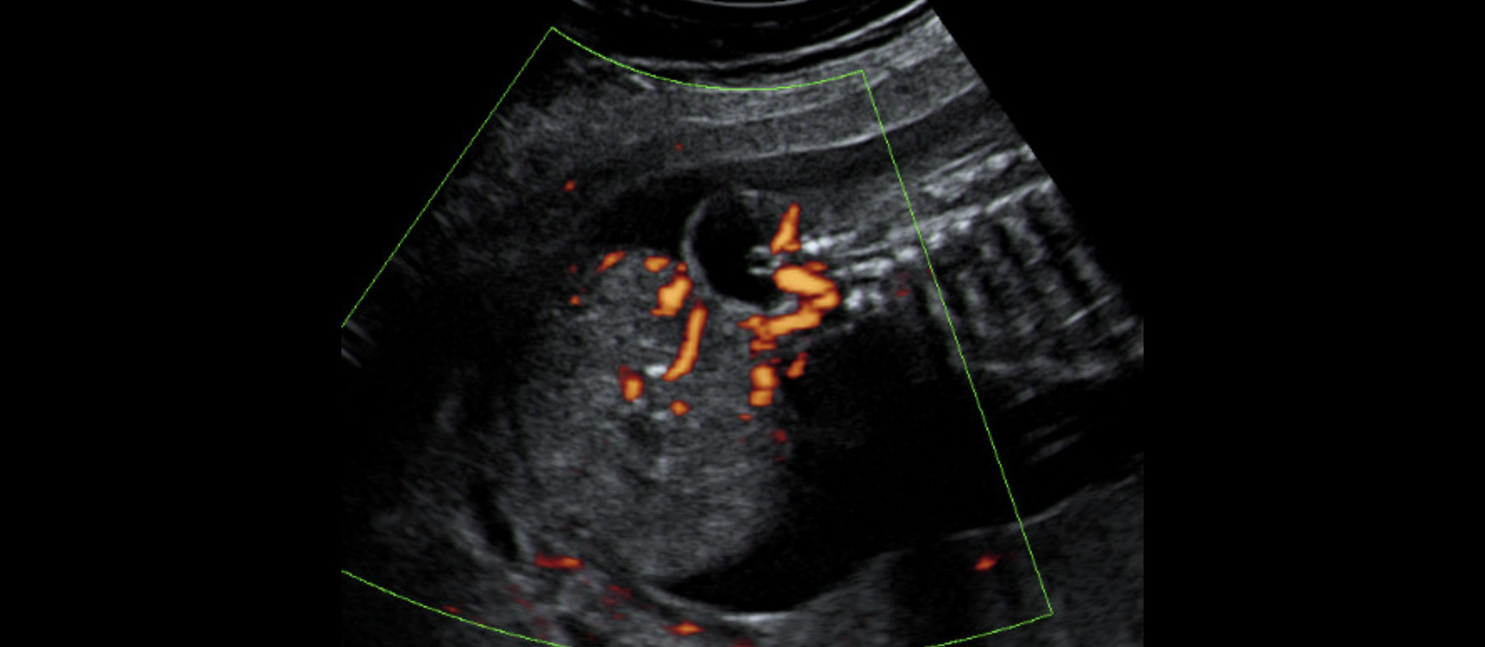
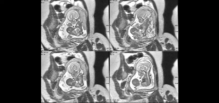
Yes, research is so important! Research allows the development of new technology and all the studies needed to confirm their safety and subsequently all their applications. Now we can make use of 3D/4D fetal ultrasound and fetal MRI to better detect those problems. In addition, fetal surgeons and maternal-fetal medicine specialists have access to improved or new technology to perform treatments even while the baby, or babies in the case of multiple pregnancies, are still in the womb.
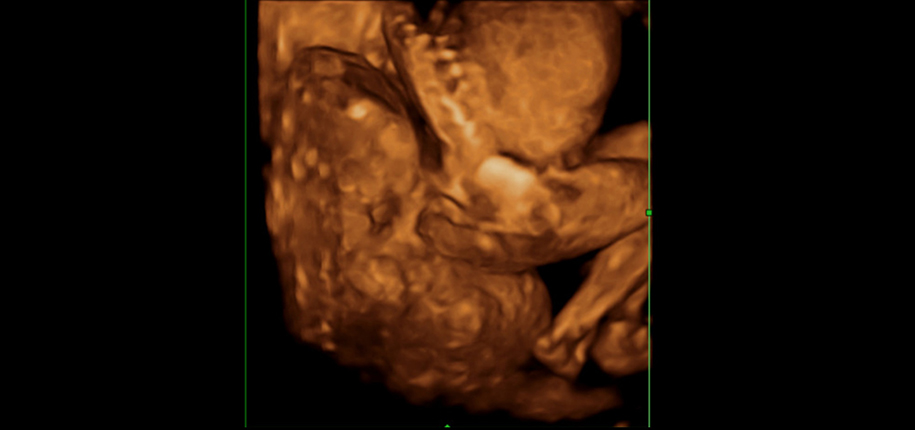
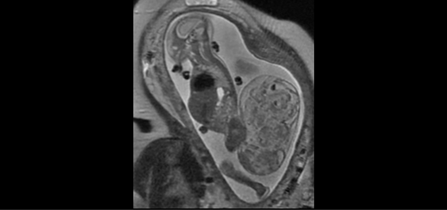
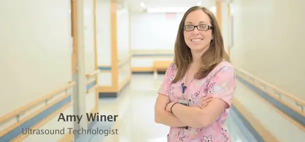
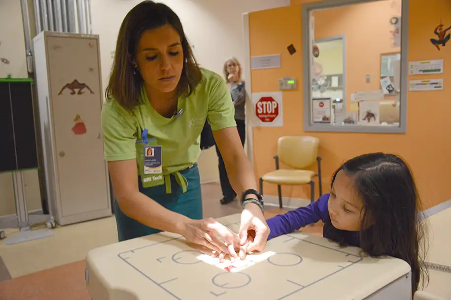
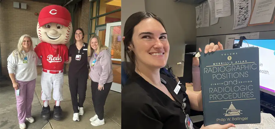
With better images, it is not only easier to make a diagnosis, but also easier to explain a problem to parents and to all medical specialists involved during counseling and planning for treatments. And how do we get those images if the babies are moving? Technology is only part of it. Our highly skilled fetal sonographers and MRI technologists are able to obtain those images with patience and knowledge. I have such a great appreciation for their work! Thanks to them we are able to obtain the most detailed information and make more precise diagnosis. They work in our department and also assist in the operating room during those surgical procedures. They help calm moms dealing with claustrophobia or other needs during their exam, they image patients in need of urgent evaluation even when the schedule is fully packed, and they work in the post-processing imaging lab obtaining measurements and calculations essential during counseling. They are key and essential members in this field that have been called Fetology, a branch of medical science concerned with the study and treatment of the baby before it is born. However, we should not forget our MRI coordinators and their diligent and efficient work behind the scenes that make it possible for all these complex imaging appointments to run smoothly even when urgencies or rearrangements are added.
The future for the field of fetal imaging will involve faster techniques with improved images that are easier to analyze. However, the spirit of teamwork and dedication among all members of the fetal imaging team will remain in the background and on the frontline, as it has always been!

Dr. Maria Calvo-Garcia, author; Glenn Miñano, BFA, editor; Meredith Towbin, copyeditor.
Glenn Miñano is a media specialist in the Department of Radiology, providing graphic design, photography, printing, video services, and administration of the department’s online properties. His works have been published in several medical articles, such as the American Journal of Radiology and the American Institute of Ultrasound. He has been providing these services to the Radiology Department since 1996.