While our #MummyScan is officially complete and the results have been shared, there remain some unanswered questions about the Peruvian child mummy on display at the Cincinnati Museum Center.
Before this winter, not much was known about this 500-year-old mummy which is part of Mummies of the World: The Exhibition, now on display at the museum center through the end of April. We were approached by the museum center and exhibition staff to perform a virtual autopsy to help shed some light on how this child lived and died.
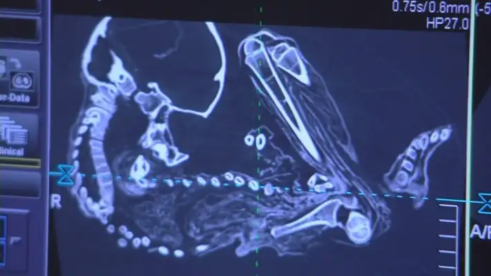
On January 28, a team of radiologists, technologists and I volunteered to image the child here at Cincinnati Children’s. We first took x-rays to get a sense of the position of the mummy and if any objects had been buried with the child. We followed the x-rays with CT scans to get a more detailed look at the bones and soft tissues and to allow us to develop 3D models.
By the time we finished the imaging and the image reconstructions, we had created more than 12,000 images. In our analysis of these images, we took the same approach that we do to evaluating the imaging of any child we might see here at Cincinnati Children’s. That approach takes advantage of the fact that we have individual team members who are experts in specific types of imaging.
For this project, I worked closely with Dr. Jim Leach, a pediatric neuroradiologist; Dr. Tal Laor, a pediatric musculoskeletal radiologist; and Dr. Alexander Towbin, a fellow pediatric body imager. We reviewed the imaging and focused on evaluating the images related to each of our own areas of expertise. About a month after the imaging was performed, we came together as a team to meet, discuss our findings, and come to some conclusions about the child’s sex, age and cause of death.
We believe this child was a female who would have been approximately two and a half years old at the time of her death. We did not see findings of chronic disease or trauma that might have caused this little girl’s death. While we know this child died naturally, we were unable to pinpoint exactly why the child died.
This project, in addition to allowing us to participate in scientific discovery with the Cincinnati Museum Center, also presented an opportunity to leverage an emerging technology here at Cincinnati Children’s. Through collaboration with Matthew Batie, a clinical engineering specialist, we created a 3D printed model of the mummy’s skeleton. This model allowed us to better visualize and understand the mummy’s position and some of the bone findings we identified on the imaging. In addition to this 3D printed model, which aided in our analysis, we also collaborated with our colleagues Dr. Ryan Moore in the Heart Institute and Sam Antoline of the University of Cincinnati’s College of Engineering and Applied Science fabrication lab to print a high resolution version of the mummy’s skull.
These 3D printed models along with images from our scans now exist for future studies and will travel with the exhibit for the public to see. Take a look for yourself at Mummies of the World: The Exhibition which runs at the Cincinnati Museum Center through April 26, or revisit the hashtag #MummyScan on Facebook, Twitter and Instagram for more information and images.
Dr. Andrew Trout is an assistant professor of Radiology and interim director of Thoracoabdominal Imaging. His areas of clinical practice include general pediatric radiology, thoracoabdominal imaging and nuclear medicine.