
Magnetic resonance imaging (MRI) has the potential to evaluate the progression of lung disease in pediatric patients. Premature infants, for example, very often experience pulmonary complications and could greatly benefit from several imaging check-ups over time. Computed tomography (CT) is used occasionally on infant patients, but there is increasing recognition that the cumulative ionizing radiation exposure from repeated CTs is non-trivial. In comparison, MRI is a completely non-ionizing imaging method and so is ideal for imaging a pediatric patient repeatedly.
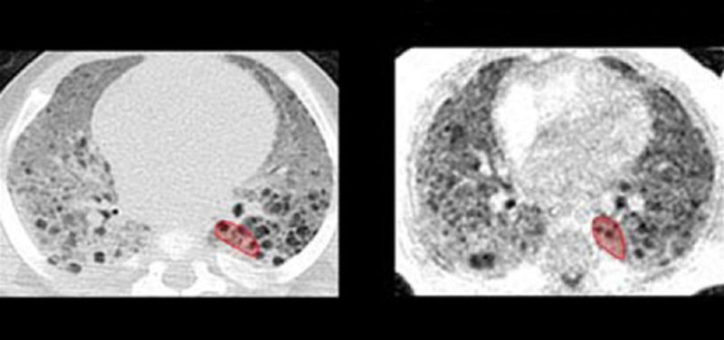
Our study on infant lung imaging uses a new type of MRI technique called “ultrashort echo-time” (UTE) MRI, which is optimal for imaging diseased lung structures. We acquired UTE MRI images from infant patients with various lung diseases who also had a clinical CT performed, so that we could directly compare image results between the two methods. With the help of a radiologist, we were able to show significant similarities between how both UTE MRI and CT represent pathological lung structures within a given patient. We were also able to show that our UTE MRI method can give quantitative measurements of lung density (in grams per cubic centimeter), just like CT can. This means that there is strong potential for UTE MRI techniques to replace the need for CT, so that we are able to repeatedly image the progression of lung disease in sick infants without worrying about the risks of ionizing radiation. Serial imaging of a patient’s pulmonary condition could help personalize their medical care in the long term.

Figure 1: Slice-matched images comparing clinical CT scans (left column) and research UTE MRI scans (right column) in five diseased subjects (from top to bottom: Subjects A through E). CT and MRI exams were typically performed at different time points for each individual subject; chronological ages at each subject’s CT and MRI exams are provided.
Quantification of Neonatal Lung Parenchymal Density via Ultrashort Echo Time MRI With Comparison to CT
Nara S. Higano, MS, Robert J. Fleck, MD, David R. Spielberg, MD, Laura L. Walkup, PhD, Andrew D. Hahn, PhD, Robert P. Thomen, PhD, Stephanie L. Merhar, MD, Paul S. Kingma, MD, Jean A. Tkach, PhD, Sean B. Fain, PhD, and Jason C. Woods, PhD
Read the full research article (click here).
Contributions by Nara Higano (Graduate Student , IRC) and edited by Glenn Miñano, BFA.