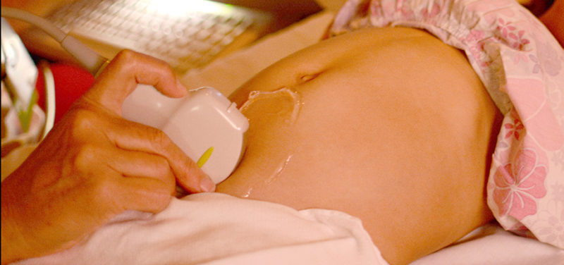

A new technology is being utilized in the Ultrasound Division here at Cincinnati Children’s: shear wave elastography. Pathological conditions can alter the elastic qualities of the tissues involved. Elastography is a non-invasive form of tissue characterization used to determine tissue stiffness. This information can aid in the diagnosis, treatment and surveillance of many disorders.
There are two main types of elastography: strain elastography and shear wave elastography. Strain elastography uses mechanical compression; the technologist manually pushes the ultrasound probe against the patient’s body part being investigated. This method results in a qualitative comparison of tissue stiffness.
Figure 1. The technologist pushes the probe against the patient.
 Image 1. The colors in the image illustrates the degree of softness or stiffness.
Shear wave elastography, however, uses a push-pulse or force that is sent out from the probe. This method provides a quantitative analysis of tissue stiffness, producing measurements relating to the stiffness of specific lesions or diffuse disease. This method is quick and painless for the patient, and can be completed within minutes.
Figure 2. The probe emits a painless push-pulse into the patient.
 Image 2. The colored lines illustrate the pulse propagating through the tissue. The circle represents the region of interest from which the quantitative data is collected.
While elastography was first used in breast imaging, its uses now include the liver, thyroid, prostate, and musculoskeletal system. New applications will continue to be investigated with this exciting new technology here at Cincinnati Children’s.
Contributed by Janet Adams and edited by Paula Bennett.