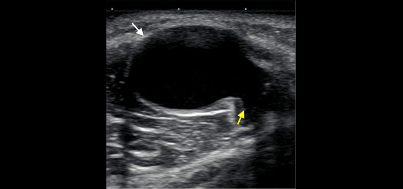

Image: Ultrasound obtained behind the knee of a young child with a soft bump shows a round cyst (white arrow) with a “tail” (yellow arrow) extending deep toward the joint between the muscles. This is a typical appearance and location for a benign cyst (also known as a Baker cyst at this location).
A newly noted lump or bump in a child is a common concern brought by parents to pediatricians. Depending on your child’s symptoms and physical exam findings, your child’s pediatrician may order an ultrasound to gather more information about the underlying problem.
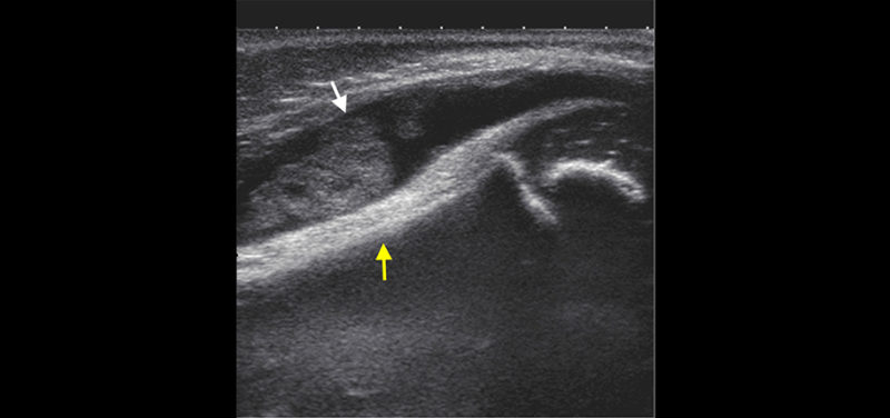
 Image: Ultrasound obtained at the front of the knee in a young child with swelling shows a fluid collection in the knee joint that contain debris (white arrow). The femur is noted by the yellow arrow. This patient was ultimately diagnosed with an inflammatory condition of the joints.
Ultrasound is a fantastic way to take a deeper look at what could be causing the issue, particularly in a young child. The advantages of ultrasound in this scenario are numerous, including:
Related Article: Ultrasounds and Bladders: What’s the Connection?
After the ultrasound technologist scans the patient and shows the images to the radiologist, the radiologist may come in to obtain more images and ask more questions about what you have noticed about the bump. Once the radiologist has finished scanning, he or she will likely be able to give you some ideas about the underlying cause for the lump. A specific diagnosis may be clear, or further testing (such as an MRI) may need to be done in the next few weeks to gather more information. Sometimes, a follow-up clinical exam or ultrasound may be indicated. Rarely, a child may need to be scheduled for a biopsy/removal of any associated mass. Regardless, the radiologist will often be able to tell you what he or she is going to report to your child’s pediatrician and may be able to give you a general idea about what to expect next from the ordering pediatrician. It is, of course, completely appropriate to ask questions of the radiologist before he or she leaves the room—we are happy to give you as much information as possible!
Contributed by Dr. Carl Merrow Jr. and edited by Glenn Miñano, BFA.
