Have you ever wondered what happens if you need an MRI but you have metal or implants in your body? Can the scan be done? That is a terrific question, but in short, the answer depends on many factors. Cincinnati Children’s MRI division prides itself in evaluating those factors to make patient safety its top priority.

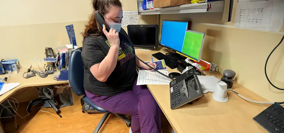
The first, and most important, step is metal safety screening. ANYONE who enters the MRI zones must be screened for metal both outside and inside their body. The MRI is a super-conducting magnet. It is ALWAYS on. Our MR technologists thoroughly screen patients and family members prior to entering the scan room. At times, our safety questions seem bothersome and redundant. For example, you may answer questions during the original scheduling call, then again during a confirmation call, yet again when you arrive and speak to one of our nurses and/or patient care assistants, and lastly when the MRI technologist meets you. These steps are deliberate to verify safety precautions. The MRI technologist makes the final decision to enter the scan room. Yet, if questions persist, the technologist may enlist the help of a radiologist or our MR safety director, Dr. Stacey Elangovan.
Once your metal safety screening is completed. We evaluate what implants pose a concern. In most cases, we research the implant and check the manufacturer’s specifications. Many implants are made of silicone, copper, carbon fiber, aluminum, or titanium, to name a few. These are non-ferrous materials- meaning they do not attract to a magnet. Most are SAFE to enter the MRI scan room. However, many manufacturers recommend that we change the settings on our scanners to use specific scan parameters when imaging the implant, so that we stay within their safety guidelines. These implants are considered CONDITIONAL. Other implants are NOT SAFE to enter an MR environment. Examples of NOT SAFE implants include most pacemakers, defibrillators, and cochlear implants.


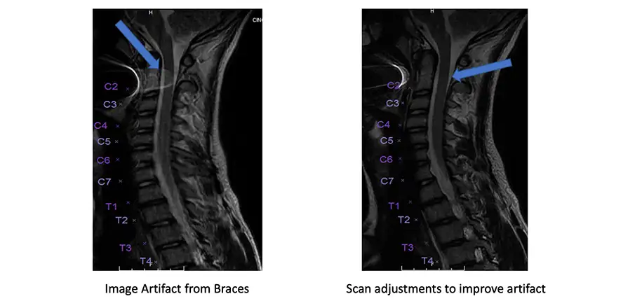
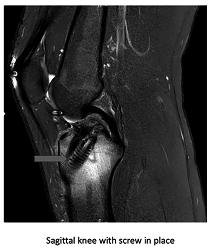
After safety steps are taken, the patient can enter the MRI scan room. When taking the pictures, sometimes the implant does not affect the image. An instance of this would be if a patient has braces on their teeth but we are scanning their knee; the braces will not affect the images because we are not scanning that body part. However, if we are scanning a patient’s spine and they have rods or screws in the back from a prior surgery, technologists and radiologists will expect to see an image artifact. Image artifact is a visual distortion of the picture. The severity of the artifact is influenced by several factors. It depends if the implant is big or small, how many there are, and what material it is made of.

When scanning with an implant, technologists use special sequences to minimize the artifact. Radiologists use the patient’s prior MRI scans and compare them to the study with distortion to make a diagnosis. In rare instances, the artifact is too great and the exam is undiagnostic. In those cases, another imaging modality like CT or ultrasound is used.
With current medical advancements, conditions are always improving. Manufacturers that make the implants are modifying the materials that they use so that they are more favorable for MRI imaging. Companies that make MRI magnets are developing new sequences to help diminish distortion from implants.
As a leader in the industry, Cincinnati Children’s applies these new techniques to provide your child with the best images possible.
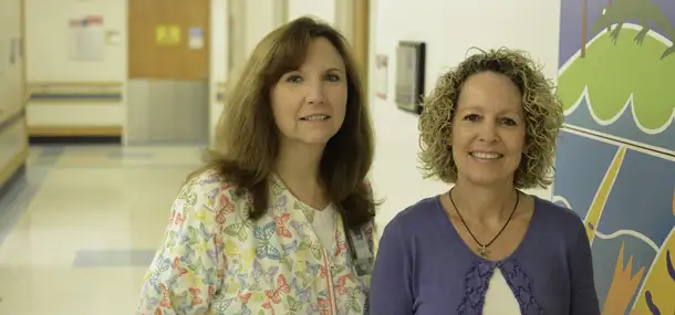
Angie has been part of the Cincinnati Children’s Radiology family since 1996. Over the years, she has worked in X-Ray, Cat Scan, and most recently in MRI. Currently, she is part of the Outpatient MRI Team, primarily working at the Liberty Township, Green Township, and Kenwood Campuses. Angie enjoys working in a stimulating environment and learning new things each day. In her free time, she stays active with her husband, two teenage daughters, and her Golden Retriever, Sonny.
Glenn Miñano is a media specialist in the Department of Radiology, providing graphic design, photography, printing, video services, and administration of the department’s online properties. His works have been published in several medical articles, such as the American Journal of Radiology and the American Institute of Ultrasound. He has been providing these services to the Radiology Department since 1996.
Editor: Meredith Towbin