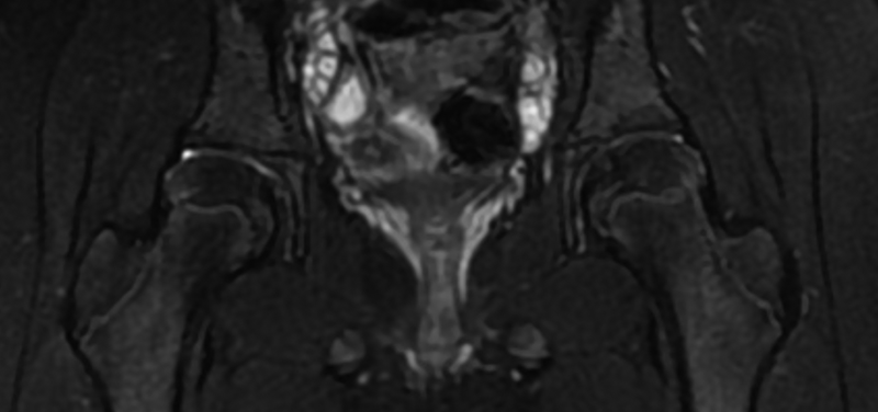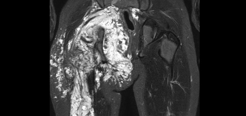
Doctors can have a hard time evaluating the pelvis. This is because its internal structures are deep and difficult to fully examine. When children have pain or other symptoms in the pelvis, doctors will often ask radiologists to help them figure out what is causing the symptoms. The Radiology Department at Cincinnati Children’s performs a number of different tests, depending on the condition or symptoms. Some of these tests include ultrasound, CT, nuclear medicine, fluoroscopy, and MRI. MRI is often the best test to evaluate for conditions that children are born with (called congenital malformations), inflammation or infection (such as Crohn’s Disease or infections of the bone), or tumors (such as neurofibromatosis or Ewing’s sarcoma).
 Image: Here is an image with a large neurofibroma of the right pelvis.
MRI is great test for certain conditions because it provides excellent detail and involves no radiation. This means MRI is good for children who need multiple exams over time in order to monitor their condition. Because MRI depicts anatomy in greater detail, radiologists can distinguish the different parts of the body that reside in the pelvis, even when they are quite small. Being able to distinguish these small body parts allow radiologists to find parts that are different than normal and then determine what effect these differences will have on later growth and development. The hardest part of MRI is that your child will have to lie still in the scanner during the 45 to 75 minutes required to take the pictures. We try to make this as easy as possible for your child by letting him or her watch a favorite movie during the scan.
Contributed by Dr. Kathy J. Helton-Skally and edited by Dr. Alex Towbin.