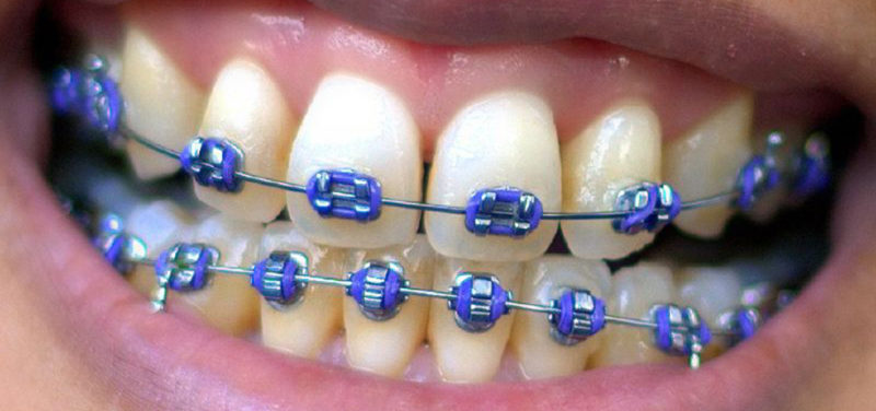
Magnetic resonance imaging, or MRI, is a non-invasive imaging exam that uses radio waves and magnetic and fields gradients to generate anatomic images of a body part. It does not use ionizing radiation like CTs or x-rays. Prior to MR imaging, the patient must be free of removable metal objects like coins and keys, as metal objects may cause safety hazards for the patient. Some metal objects cannot be easily removed, like braces or implantable medical devices. Because of this, some patients may not undergo MR imaging because of the risk of damage to the devices or heating resulting in damage to surrounding tissues.
However, patients with many types of orthodontics, including metal braces, retainers and some palatal expanders, may undergo MR imaging, as these are sufficiently secured to the teeth and have been shown to be safe on clinical MR scanners at 1.5T and 3T (tesla). Even though they may be safe, these orthodontic devices may cause artifacts to occur on the generated images. These artifacts occur predominantly in the facial and orbital regions, although artifacts can also extend to include areas of the brain and cervical spine.
 Image: Ax T2-weighted image shows artifact from orthodontics partially obscuring the face (red arrows).
Even with artifact resulting from the metal orthodontics, very few patients will need to have the orthodontics removed to obtain adequate imaging. Different MR techniques and imaging angles can aid with alleviating some of the artifact and provide more optimal images. Rarely, a patient may need to have an orthodontic device removed to allow the radiologist and patient’s physician to closely follow a lesion or evaluate structures near the face that are obscured by the orthodontics. Also, uncommonly, a patient may be asked to have their orthodontist remove a specific device to allow a patient to safely undergo MR imaging without the concern of injury to the patient. However, removal should be performed only after discussion between the ordering physician, patient or parent, and the radiologist.
 Image: Ax susceptibility-weighted image shows extensive orthodontic artifact obscuring the anterior brain (blue arrow). On the T2-weighted image in the same patient, the brain is well seen (yellow), and the artifact is not a factor.
Contributed by Dr. Marguerite Caré and edited by Michelle Gramke, (Adv Tech US).