As you probably have come to expect from Cincinnati Children’s Radiology Department, we are constantly trying to push the envelope everyday and in every way: from the way we tackle new and improved technologies to how we go above and beyond to accommodate our patients and their families. A recent grand example of this can be witnessed when visiting the MRI Department’s cardiac unit.
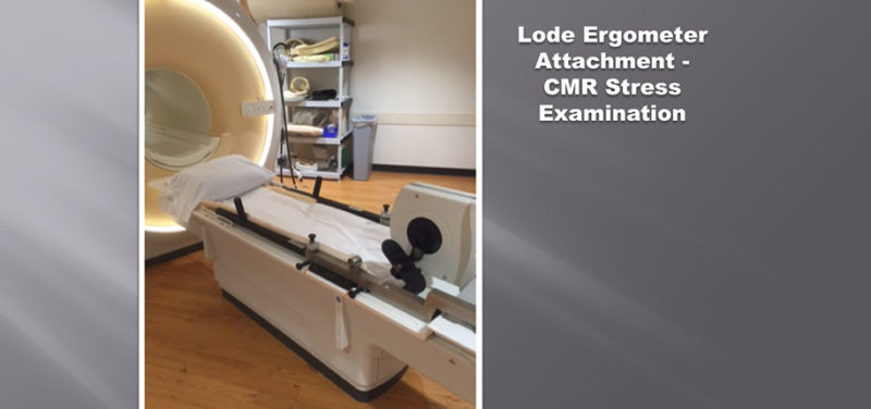
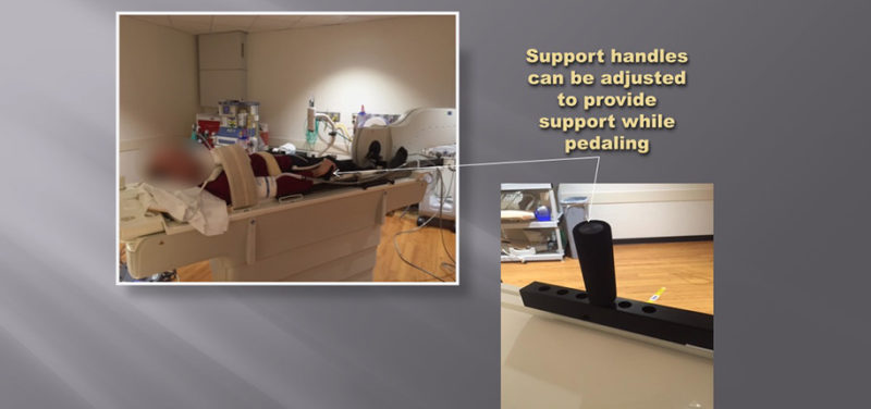
Not only do we have an MRI scanner devoted primarily to cardiac studies, but we also strive to adopt new equipment to image cardiac diseases in innovative ways. This is why our cardiac MRI on A6 has acquired an MR ergometer. The MR Ergometer is an alternative to the traditional pharmacological stress tests used to evaluate cardiovascular issues such as coronary disease, ischemia, heart function and flow. The unit is MRI safe and can be easily attached to the end of the MR table. The device consists of a pair of bicycle pedals and is used to simulate exercise. While laying on his or her back on the MR table, the patient pedals the bike at increasing resistance levels until an optimal heart rate is reached. Images are then taken and compared to pre-stress images. A gadolinium-based contrast agent is often used to help highlight dysfunction or ischemia following the use of the ergometer. This alternative method for stressing the heart presents less risk than other alternatives with comparable results.

In the past, the only other imaging option was to use an artificial stressing caused by giving a pharmaceutical through an IV. This medicine would increase the heart rate, like the patient just exercised, but would not show the true effects of exercising stress on the heart, much like how caffeine may keep you awake, but getting a good night’s sleep is the only way to accurately replenish alertness.
With the use of the MR ergometer and newer MRI imaging techniques we are able to view the heart in an innovative manner. As technology grows and the research for MRI-compatible systems continue to be invented, the world of pediatric imaging grows along with them, enabling us to witness effects otherwise unseen. You can bet with each invention created Cincinnati Children’s Radiology Department will be right there partnering with these new studies along the way.