Technology is advancing, and more and more often we are able to track disease with imaging, thereby avoiding biopsy. One relatively new imaging technique that we are very excited about it shear wave elastography (SWE). SWE is done with an ultrasound machine and is a way of measuring how hard tissue is. The major use for SWE is looking for and following liver disease since both swelling and scarring make the liver harder.
At Cincinnati Children’s we use Toshiba ultrasound machines, which are able to do SWE. While there is a lot of data about how very diseased livers look and measure on SWE, there is very little data about how healthy pediatric livers look and measure on SWE, particularly Toshiba SWE. Knowing exactly what normal livers look like will help us be more confident about who is healthy and should help us diagnose liver disease at an earlier stage. A current two-part study here at Cincinnati Children’s should give us a lot of data about Toshiba SWE and what healthy and diseased livers look like.
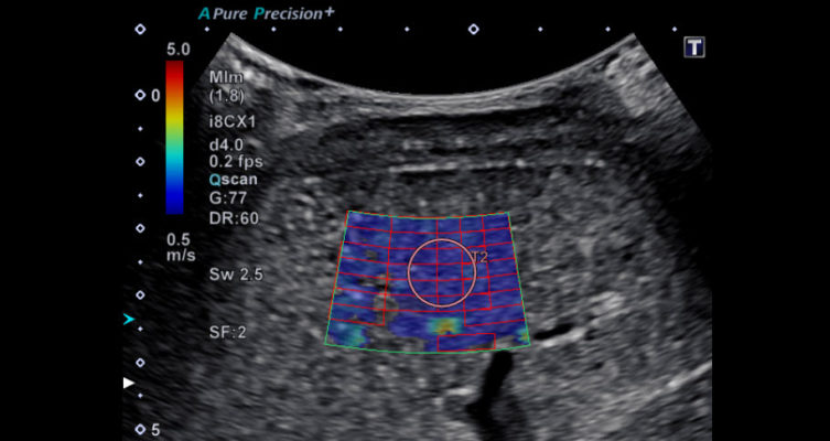
 Image: Normal shear wave elastography ultrasound in a healthy patient.
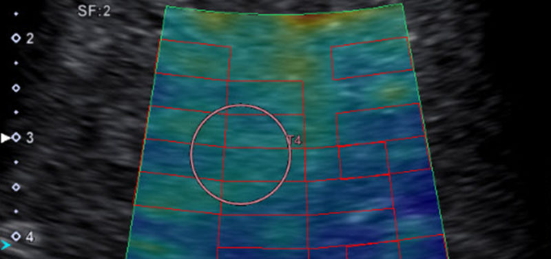
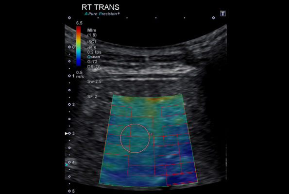
 Image: Abnormal shear wave elastography ultrasound in a patient with liver disease. In the patient with liver disease, the liver is harder with more yellow and green in the color map compared to the uniform blue in the color map from the healthy child.
Part one of this study is designed to measure normal livers with SWE in heatlhy children and a few adults. We’re also measuring normal pancreas in the same patient to see if maybe going forward we can use SWE to diagnose pancreatic disease. Part two of the study is focused on patients with liver disease, comparing ultrasound SWE to other measures of liver disease, including MRI. While we are focused on SWE in this study, we are also looking at some newer ultrasound imaging techniques to measure changes in liver tissue that might relate to early swelling and accumulation of liver fat.
Contributed by Dr. Andrew Trout and edited by Sparkle Torruella (COORD-REG AFF).
