
At Cincinnati Children’s, we perform ultrasound and magnetic resonance imaging (MRI) to evaluate babies that have been diagnosed with a problem in the womb before birth. Our Fetal Care Center is one of the most innovative in the country, providing surgical and medical interventions that can change the outcome for these babies before or at birth.
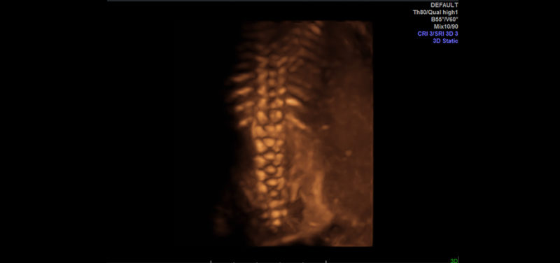
 Image: Ultrasound 3D image of fetal spine
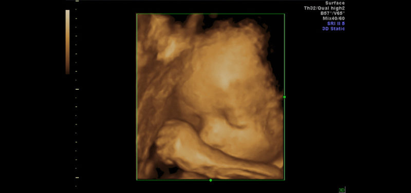
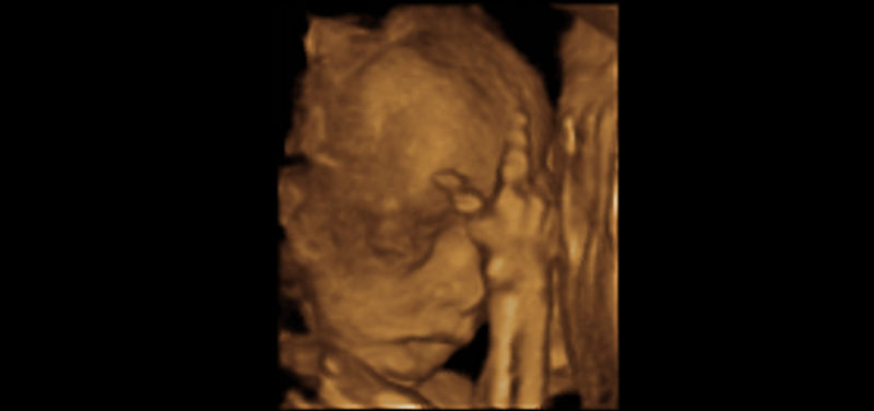
 Image: Ultrasound 3D image of fetal face
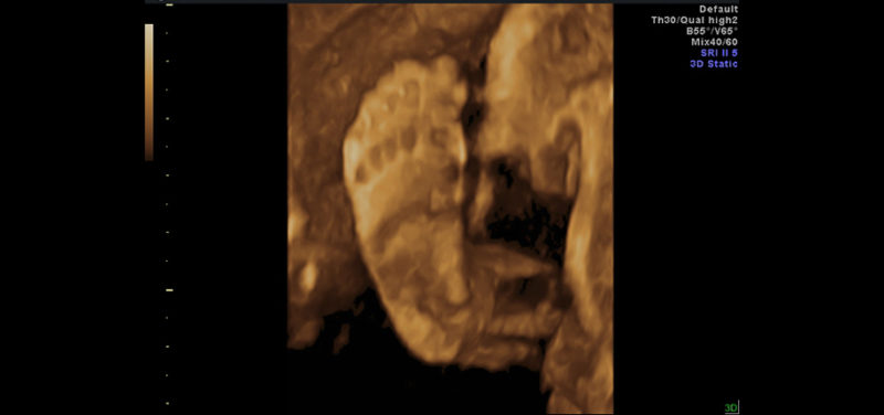
As fetal imagers, our goal is to provide as much information as possible to understand the abnormality in the baby. Ultrasound is an excellent technique to image the fetus as it uses sound waves instead of radiation to obtain the pictures. Ultrasound is exceptional at seeing soft tissue, bones and blood vessels. With 3D ultrasound, we can reconstruct those pictures into a three-dimensional image to see the baby’s face, bones in the skull or spine, or arms and legs. With these images, we are able to provide more information about abnormalities. This additional detailed information is helpful in counseling the parents and preparing for surgical intervention before or after birth.
 Image: Ultrasound 3D of fetal foot
Our imaging modalities, especially ultrasound, continue to support our Fetal Care Center so that the best care can be provided for the babies referred to our hospital. With the amazing images we can obtain of the baby before birth, we are working hard to improve the outcome for our tiniest of patients and their families.
Contributed by Dr. Beth Kline-Fath and edited by Janet Adams, (ADV Tech-US).
