Every year in the United States there are approximately 1,750 spinal cord injuries in children and teens younger than 18 years of age. Here at Cincinnati Children’s, my colleagues and I are interested in discovering early imaging signs of traumatic injuries that may lead to spinal cord damage if not properly treated. This type of research has been done in adults, but the spines of children are very different from adults and change (grow) dramatically from birth to teenage years.
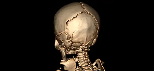
Our research first focused on defining normal bony measurements of the joint that connects the skull to the spine, which is called the occipital condyle-C1 interval (or CCI for short). Using CTS scans of this joint we found that children have a much larger bony measurement than adults because their bones will not completely develop until their late teenage years.
The second part of our research compared these normal CT joint measurements to the many cases of spine injury that we have seen over the years here at Cincinnati Children’s. We found that many of these patients with injuries at the joint between the neck and head had abnormally large joint measurements, suggesting dislocation. Unfortunately, there were some patients with injury that had normal measurements, telling us that CT alone is not enough and sometimes MRI is necessary to more confidently make the diagnosis of injury. Luckily Cincinnati Children’s Radiology Department makes it very convenient for you and your family to rotate from one imaging area to the next, allowing you to easily acquire all your imaging needs under one roof.

Contributed by Dr. Luke Linscott and edited by Tony Dandino, (RT-MR).
Tony is an MRI Technologist at Cincinnati Children’s. Tony has been in his role for several years and serves as a Charge Tech, Quality Improvement Coach and Safety Coach for the MRI department. Tony has always known he wanted to work with children and in the medical field. Working at Cincinnati Children's has been the best of both worlds. Every day is something new and Tony can never wait to start the next adventure.