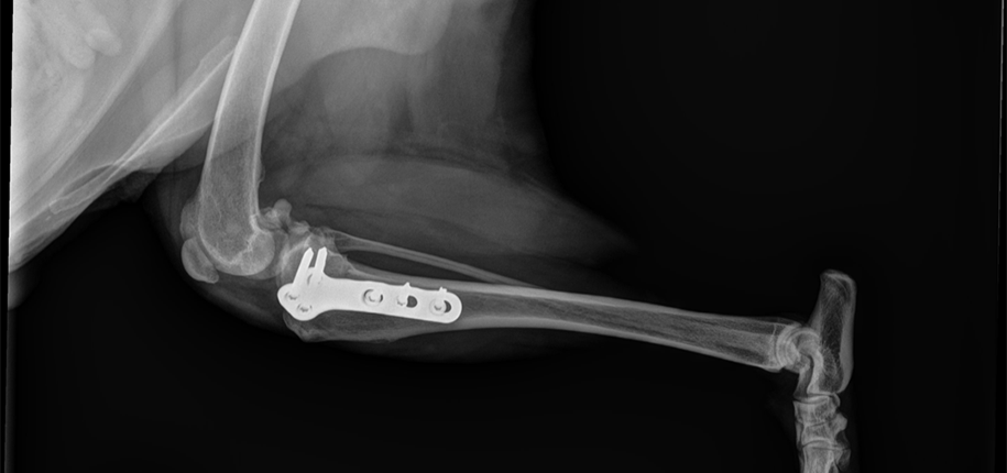
As a Pediatric Radiology Fellow, I spend my days interpreting images, including x-rays and ultrasounds on children of all ages. X-rays create a picture of the bones and joints that help us find fractures and other abnormalities. They are an important diagnostic tool in human medicine, but not many people know that x-rays are just as important in animal medicine. This is how x-rays helped save my dog’s leg.

Recently, my dog Bear was avoiding going outside to play. When I examined him, he was holding up his right leg, not wanting to move. Normally he is very active and loves to (playfully) jump on the trees to try to catch squirrels. Although the squirrels were happier, I was pretty worried and took Bear to the veterinarian.
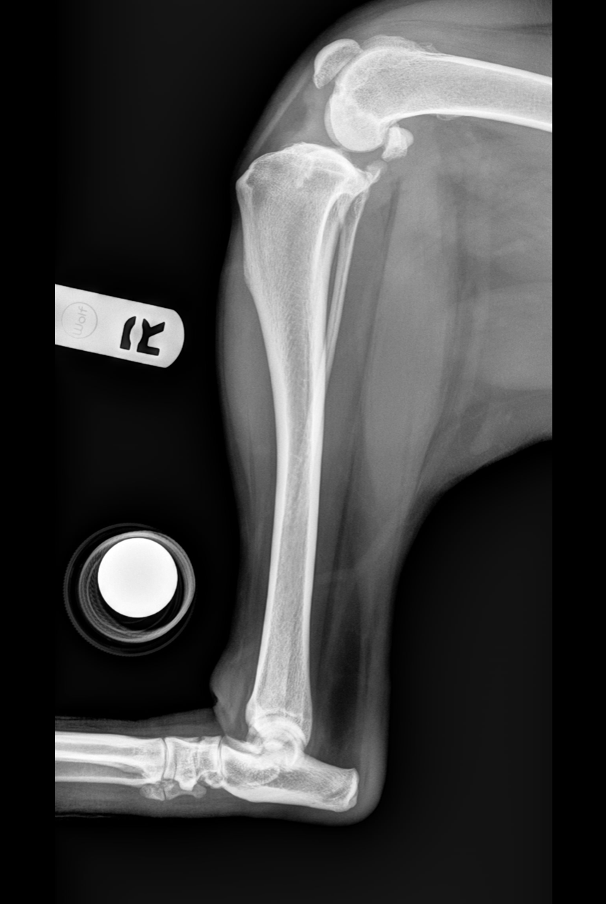
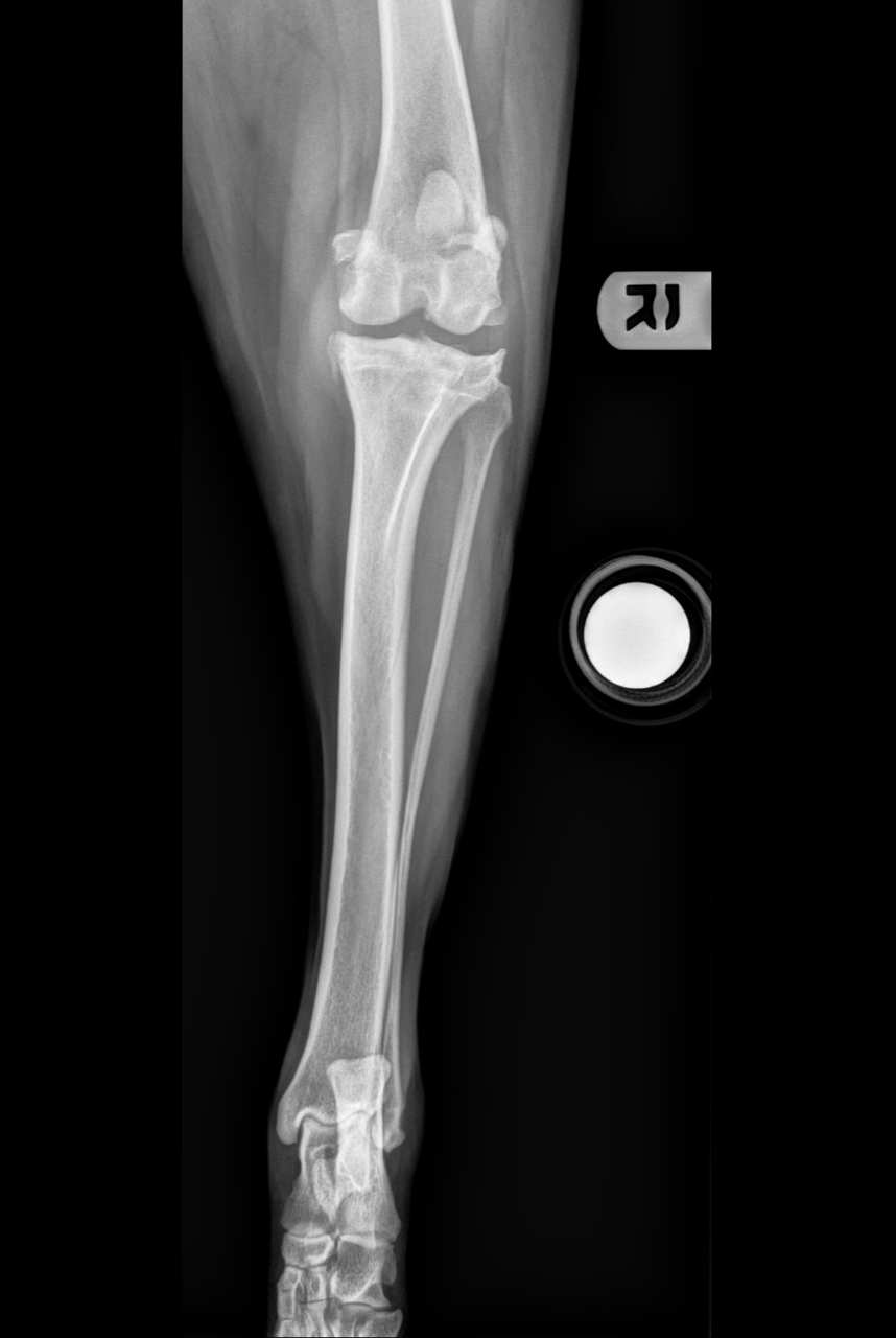
Bear’s doctor immediately took an x-ray. To me, this x-ray looked vaguely familiar, as dog bones look a lot like human bones. Based on only the x-ray, Bear was diagnosed with a tear of his canine cruciate ligament, which normally provides stability to his knee joint. This is very similar to an anterior cruciate ligament (ACL) tear, a common injury that famous athletes get. Radiologists for humans usually diagnose ligament tears using MRI, which provide 3D images. It’s fascinating that animal doctors can make this diagnosis using only x-rays!

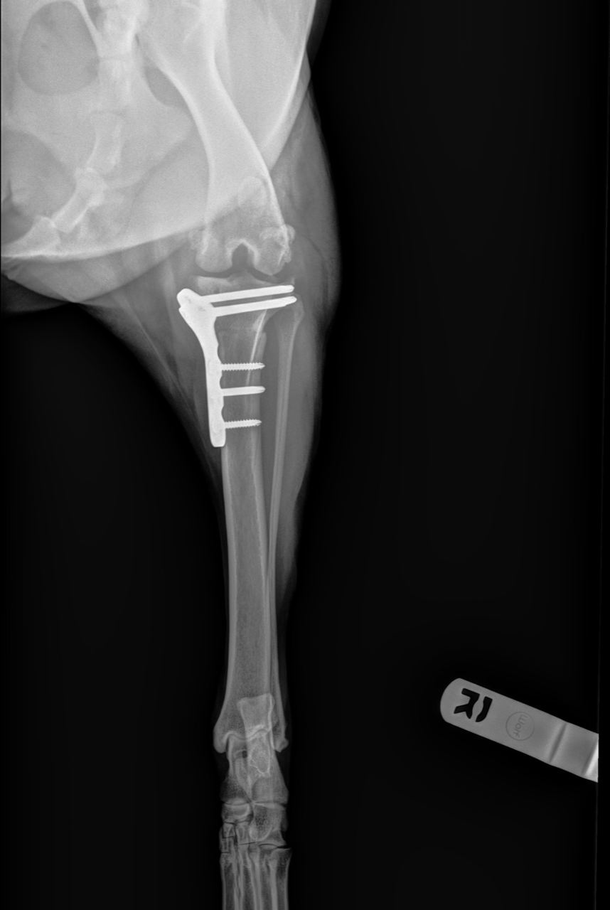
To help Bear walk and prevent pain, he had to have a complicated surgery on his knee. X-rays were also used during and after surgery to make sure the surgical hardware was okay and the bones were healing. This is very similar to what we do with our patients here at Cincinnati Children’s when they have a broken bone requiring surgery. Radiology can help both humans and animals!
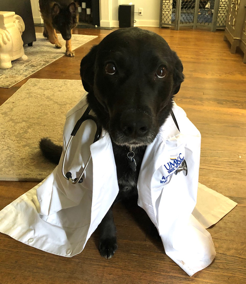
For those wondering, to the squirrel’s dismay, Bear is back up and jumping!
Dr. Joshua Wermers, author; Glenn Miñano, editor; Meredith Towbin, copyeditor.