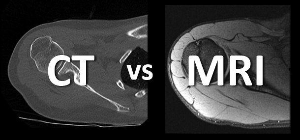
When I started experiencing shoulder dislocations due to a fall, the first test that was ordered was an MRI and then later a pre-surgical CT. I started wondering: What’s the difference? Why couldn’t the MRI images be used for pre-surgical review?
What I found out was that MRI and CT scans both provide diagnostic images, but each exam is ordered based on what you can see more clearly on the images. In some cases, both may be needed.
 Image: MRI of the shoulder shows how different parameters on the MRI scanner can make the images look different. Radiologists use these differences to determine what is wrong.
MRI scans are best suited for looking at soft tissues such as ligaments and tendons, the brain, and many of the internal organs. MRI can provide exquisite detail of these structures and allows radiologists to diagnose a wide variety of disorders such as injuries to tendons and ligaments, strokes, and tumors. Radiologists design each MRI scan to answer the specific question your child’s doctor is asking. In some cases, MRI can even determine how your child’s body works and provide information on the cells that make up his or her body parts. An advantage of an MRI is that there is no radiation used, but the scan takes longer than a CT (30 minutes or longer).
 Image: CT of the shoulder shows the detail in the ball and socket joint of the shoulder. Radiologists used these images to make the three-dimensional picture of the bones.
CT scans are very quick and provide a different level of detail. CT does not have as many options for the radiologists to select, so the scan can usually be completed in 10 minutes or less. The actual time the CT scanner is working is usually less than 10 seconds. CT is better than MRI in looking at the lungs, detecting certain bone injuries, and identifying urgent issues throughout the body. One advantage of CT is that radiologists can use computers to create very detailed three-dimensional models of the structures they are seeing.
 Image: Head CT and brain MRI show some of the differences in detail between the two different tests.
In the case of my shoulder injury, the MRI was used by the radiologist to determine what was wrong (torn labrum). The CT was used to show the detail in the ball and socket joint of the shoulder. This information was needed to determine the amount of bone loss to determine what type of shoulder surgery was needed.
Images contributed by Dr. Alexander J. Towbin.