Obesity is a major public health problem, and obesity among children is becoming increasingly common. Obesity can impact multiple body organs and systems and is associated with other diseases such as diabetes. One of the organs that is particularly impacted by obesity is the liver. As we gain weight, fat can collect in the liver where it can cause irritation and scarring. Non-alcoholic fatty liver disease (NAFLD) is the general term for liver disease due to fat depositing in the liver and is becoming a major cause of liver disease in both children and adults. Here at Cincinnati Children’s, we have clinics that care specifically for children and young adults with suspected NAFLD.
In the past, liver biopsy was the only way to accurately identify and measure fat collecting in the liver. New imaging techniques are beginning to replace, or at least supplement, liver biopsy. MRI is ahead of all other imaging techniques in terms of identifying and measuring liver fat without a biopsy. With one breath hold, we can now quantify, with good accuracy and repeatability, the amount of fat that is in the liver.
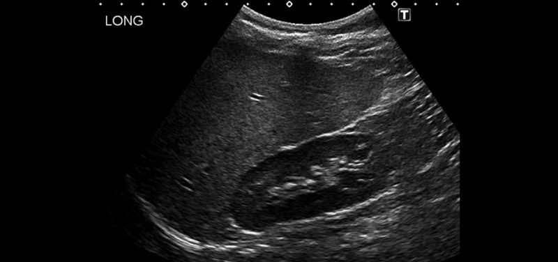
 Image: Ultrasound image in an 8-year-old obese patient shows the liver to be brighter than the kidney due to diffuse fat.
Here in the Cincinnati Children’s Department of Radiology, we are partnering with the companies that make imaging equipment and software to develop and improve imaging techniques aimed at identifying and measuring liver fat. We have already done work to show the repeatability of liver fat measurements by MRI. MRI isn’t for everyone, though, and more patients get liver ultrasounds than liver MRIs because ultrasound is more available and generally less expensive, particularly in other parts of the world. For this reason, we are now working with industry partners to test ultrasound technology that may be able to identify and quantify liver fat. Relatively soon we will be kicking off a study that will compare MRI and ultrasound for the identification and measurement of liver fat in about 50 children and young adults with NAFLD.
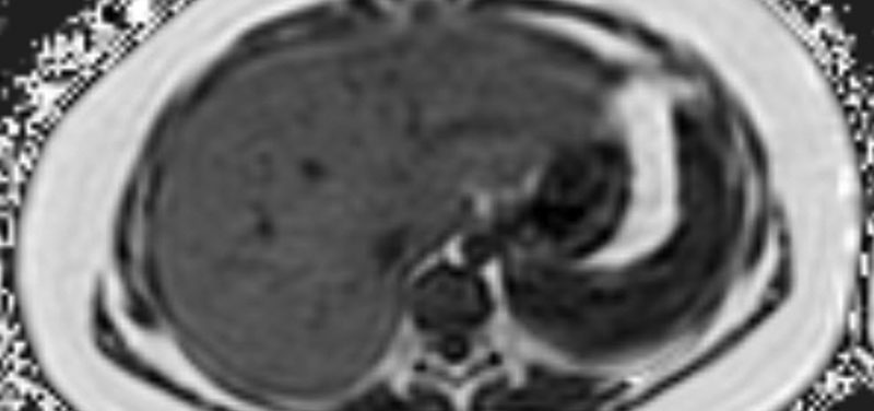
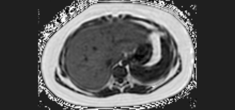
 Image: MRI fat map in the same patient shows higher levels of fat as brighter shades of white. The liver is whiter than it should be and measured to be 31% fat (normal is less than 5%).
Our hope is that by continuing to partner with industry to advance imaging science and technique, we can provide better care to more children in Cincinnati and around the world.
Contributed by Dr. Andrew Trout and edited by Sparkle Torruella, (COORD-REG AFF).
