If a child presents with an asymmetric head shape, the pediatrician may be able to determine if the child has positional molding with normal cranial sutures, which may resolve spontaneously or with positional management of the head. However, in some cases of an abnormal head shape, the pediatrician may be concerned that there is premature closure of the cranial sutures, or craniosynostosis, which may require surgical correction.
Skull radiographs or head CT may be utilized in cases where craniosynostosis is a concern. If a head CT is performed, post-processing allows generation of 3D reconstructions of the child’s skull without additional imaging or radiation exposure to the patient. These images allow the radiologist and surgeon to optimally visualize the child’s head shape and cranial sutures, as well as aid with potential surgical planning. Shaded-surface display 3D images are generated that show the bones from the “outside,” while maximum-intensity projections (MIP) are displayed as though the radiologist is looking at the skull from the inside of the head. These 3D reconstructions also allow the physicians to show the parents more clearly the imaging findings and concerns.

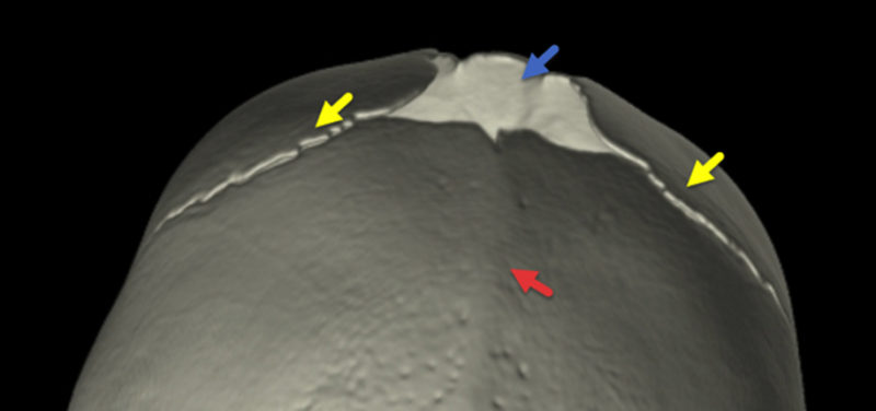
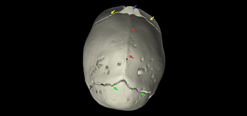
Figure 1. 3D CT reconstruction of the top of the head shows premature fusion of the sagittal suture (red arrows). The lambdoid sutures posteriorly (green) and coronal sutures anteriorly (yellow) are patent, as is the anterior fontanelle (blue arrow) or child’s soft spot.

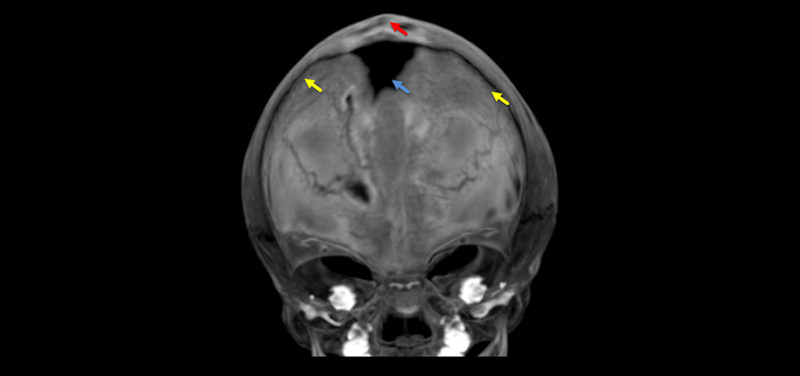
Figure 2. Maximum-intensity projection of the anterior skull again shows the fused sagittal suture (red arrow), patent coronal sutures (yellow), and the open anterior fontanelle (blue).
Contributed by Dr. Marguerite Caré and edited by Michelle Gramke,(ADV-US-Tech).
