
When I was a child, I was diagnosed with a brain tumor. Over the course of many treatments, I had a number of MRI scans to keep track of how the treatments I was receiving were working. I continue to receive MRI scans occasionally in adulthood to make sure there are no long-term complications from my treatments.
As a kid getting an MRI once every couple of months, I always had certain questions about the MRI machine. I bet kids getting scanned today have the same questions I did, so I’ll do my best to answer them!
What does MRI stand for, and how does it make images of my body?
MRI stands for magnetic resonance imaging. MRI relies on creating and changing a magnetic field in your child’s body, then measuring how certain particles in the body react to that magnetic field by using radiofrequency waves. Different tissues react differently—muscle responds differently than fat, which responds differently than bone, and so on. We take advantage of these differing properties of different tissues to create images of the body. While MRI does involve the use of radiofrequency waves, like the waves used for the broadcasting of radio stations, MRI does not involve ionizing radiation exposure like those used with CT scans or x-rays.
Why does the scanner make those loud sounds?
The large “magnet” is a coil (loop) of wires located inside the scanner and has electricity flowing through it. This main magnet creates an even or uniform magnetic field within the magnet. However, to change the magnetic field to create images, smaller magnets, called gradient coils, located within the scanner are constantly turning on and off by having electricity pumped through them. This causes the coils to vibrate as they change the magnetic field. These vibrations are the loud sounds you hear during your scan. Luckily, we have headphones inside the scanner to block out some of the sounds, which makes it easier to tolerate.
Why does the technician keep telling me to hold still or hold my breath?
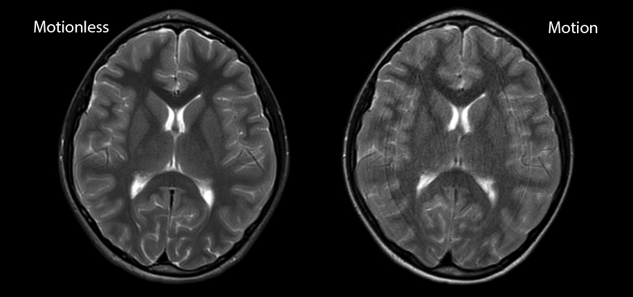
To get high–quality images of the body, we need your child to stay still. Any motion, such as voluntary or involuntary body movement, movement during breathing, or even motion related to the heartbeat, will cause what we call “motion artifact.” Here is an example:
The image on the left was obtained with the patient staying perfectly still. On the right is an image obtained while the patient took breaths freely. You can see the blurring of all imaged structures. Motion artifact makes certain things harder or impossible to see.
What is “contrast” and how does it help?
Contrast material is a liquid injected through an IV that helps us see certain structures better. Contrast material is injected directly into the bloodstream, so it goes where the blood goes. Structures inside the body that receive more blood flow than others will receive more contrast and therefore appear brighter on our images after contrast material is injected. Use of contrast material is just another tool we use to make structures in the body appear different from one-another, allowing us to tell them apart.
Dr. Nicholas Thomas-Bock, author; Glenn Miñano, BFA, editor; Meredith Towbin, copy editor