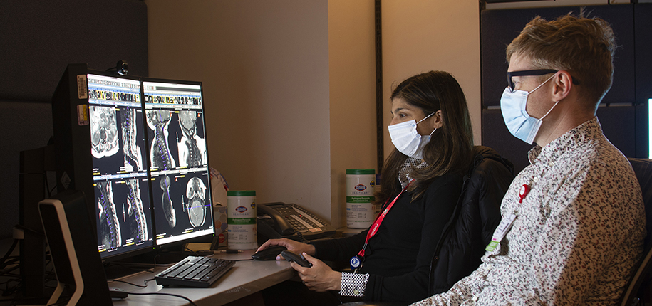
How are your images shared for patient care use? Gone are the days of darkrooms, developing film, dictated reports that are manually transcribed by office staff, and waiting for films or reports to be sent through the mail. Technological advancements are making it easier to share images and reports from institution to institution. This week, we caught up with Laurie Perry, lead systems analyst of the Radiology Informatics/Enterprise Imaging team, to explain the role of Informatics and how images are exchanged.
Because Radiology produces “images” of the body, our mantra is “image is everything.” But what does it take behind the scenes to support our vast infrastructure of images and data information? It takes a diligent, successful Informatics team. Healthcare Informatics is the field of computer science that develops methods and technologies for gathering, processing, and evaluating patient data to improve knowledge and communication within the healthcare system. Since the beginning of the millennium, our Radiology Informatics team has been responsible for overseeing Radiology workflow by supporting many internal systems including PACS (Picture Archiving and Communication System), the Enterprise Archive/Viewer and the Radiologist Voice Dictation System.
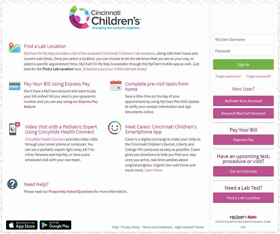
In more recent years, electronic image exchange has become an additional part of the daily workflow. This involves the transfer and sharing of images from hospital to hospital. Images were once burned to discs and taken back and forth between doctors and offices, but not anymore. According to Laurie, most large medical centers have subscribed to an image-sharing cloud service that allows them to send and receive patient images in a secure gateway. Our department generates over 224,000 exams a year. Last year, we shared/exported more than 58,000 exams to other facilities. How many studies do you think are imported into our system? A massive 95,000 exams were imported last year to Cincinnati Children’s Radiology Department. Speaking of sharing, did you know that we share your images with you on MyChart? Within 24 to 48 hours of your Radiology exam, MyChart gives you the radiologist’s full dictated report as well as a link to view your images. It’s not every day that you get to see your insides! Your child will be delighted to see his/her pictures. Be sure to take full advantage of this opportunity, as Cincinnati Children’s is one of the few facilities to offer image viewing. Click the following link for instructions to view your images on your computer or smartphone via MyChart.
https://mychart.cincinnatichildrens.org/MyChart/en-US/docs/viewing-images.pdf


Angie has been part of the Cincinnati Children’s Radiology family since 1996. Over the years, she has worked in X-Ray, Cat Scan, and most recently in MRI. Currently, she is part of the Outpatient MRI Team, primarily working at the Liberty Township, Green Township, and Kenwood Campuses. Angie enjoys working in a stimulating environment and learning new things each day. In her free time, she stays active with her husband, two teenage daughters, and her Golden Retriever, Sonny.
Angela Asher, RT (R)(CT)(MR) author; Laurie Perry, Lead Systems Analyst, contributor; Glenn Miñano, editor; Meredith Towbin, copy editor