When a sick child presents to a clinic or emergency department, the doctors and nurses begin collecting information about the patient to formulate a differential diagnosis, which is a list of reasonable possibilities for what could be causing the patient’s problems. As more information is collected, by reviewing the patient’s symptoms, physical exam, laboratory work, and imaging studies, that list of possibilities is gradually narrowed until the diagnosis is clear, which then allows the appropriate treatment to be instituted.
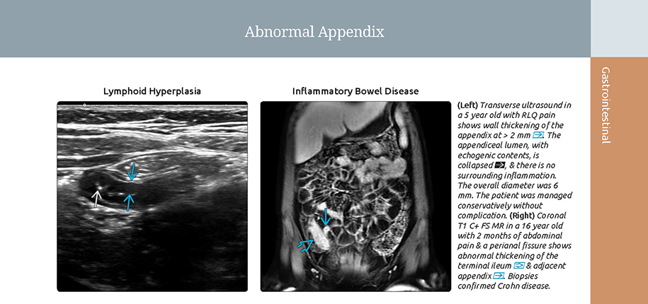
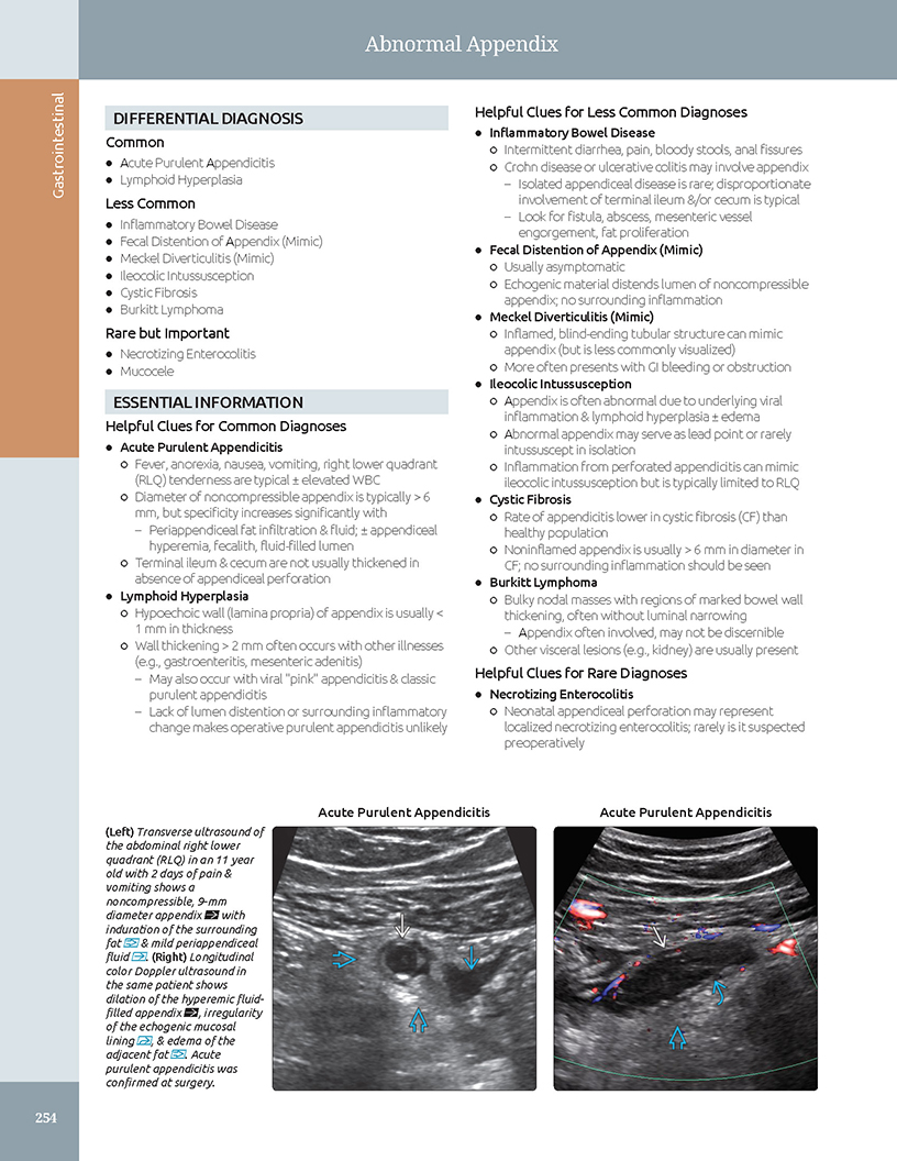
Radiologists play a critical role in this assessment. When the clinical doctor orders an x-ray, CT scan, ultrasound, or other imaging-based study, the radiologist must also formulate a differential diagnosis of considerations to explain the abnormal findings on the images of those exams. For example, an abnormally dilated appendix is most commonly due to an acutely infected appendix, but there are other less common possibilities that could create a similar imaging appearance. The radiologist must help the ordering pediatrician understand how likely those considerations are and what further evaluation can be done to come to the correct diagnosis and treatment plan.
As the editor and lead author of the newly released book Expert DDX: Pediatrics, 2nd edition, I have worked to create a text that explores the differential diagnosis of abnormal imaging findings in pediatric patients, discussing various diseases that can create similar appearances on ultrasound, MRI, and beyond. It is intended to guide radiologists to the right diagnosis of your child’s illness based on specific disease characteristics visualized on the imaging studies.
This book has been several years in the making, and I am privileged to have led an excellent team of authors in creating a reference that will be used to help children get well as soon as possible.


Contributed by Dr. Carl Merrow and edited by Glenn Miñano, BFA.
