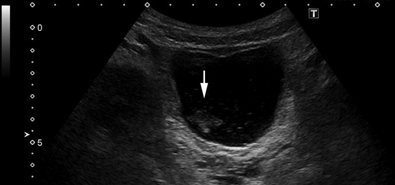
Featured Image: Large amount of echogenic debris within the urinary bladder compatible with history of urinary tract infection.
Urinary tract infections are a common problem in children. According to the American Urological Association, pediatric urinary tract infections account for more than 1 million doctor’s office visits in the United States and affect 3% of children each year. Urinary tract infection may present with classic symptoms such as urinary frequency, urgency, pain while urinating, or blood in the urine. The presenting symptoms, especially in younger children, may also be vague, such as fever, a lack of energy and enthusiasm, or irritability.
Urinary tract infections include lower urinary tract infections, or infection of the bladder, and upper urinary tract infection, or infection of the kidneys also called pyelonephritis. While it is possible for the kidneys to become infected by bacteria from the blood, many upper urinary tract infections result from bacteria sloping or leading upward to the kidneys from the bladder. It is important to recognize recurrent upper urinary tract infections because repeated episodes can damage the kidneys and lead to long-term problems such as kidney failure and high blood pressure.
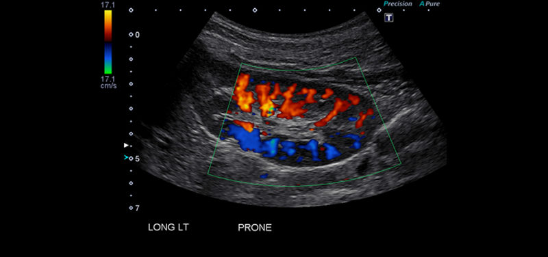
 Image: Ultrasound image of left kidney with color doppler of normal blood flow.
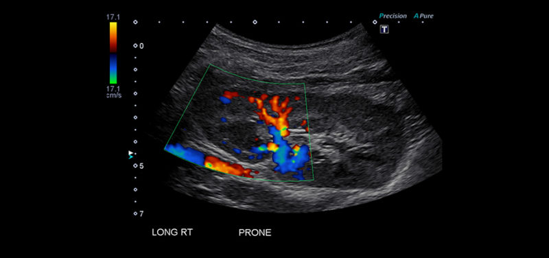
  Image: Asymmetrically enlarged and echogenic right kidney with a region of relative decreased vascularity in the upper pole; findings are suggestive of pyelonephritis.
Medical imaging plays an important role in recognizing the causes of repeated upper urinary tract infection. The American Academy of Pediatrics currently recommends that all children from the ages of 2 months to 2 years of age with a first time urinary tract infection with fever have an ultrasound of their kidneys and bladder. If the ultrasound reveals an abnormality, such as scarring of a kidney or a kidney that is dilated by urine, or if the patient has a second urinary tract infection accompanied by fever, a voiding cystourethrogram is indicated.
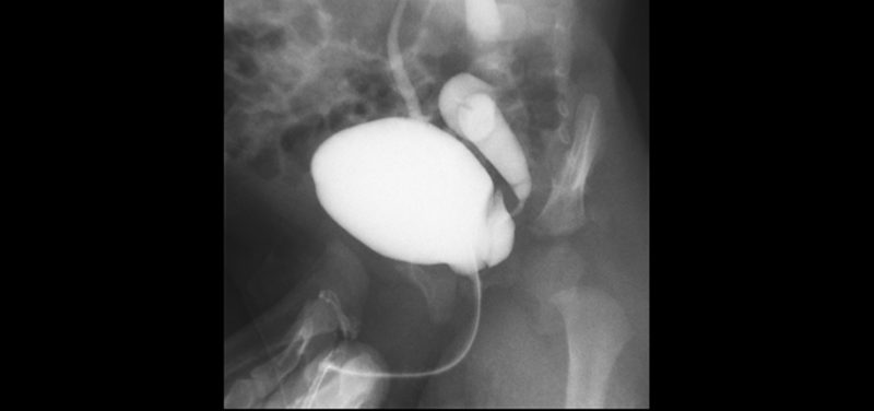
 Image: X-ray image of voiding cystourethrogram.
A voiding cystourethogram is an exam that’s performed by a radiologist under x-ray in real time. A small tube is inserted through the patient’s urethra into the bladder. Contrast that is visible under x-ray is then used to fill the bladder. Once the patient’s bladder is full, his or her urethra is observed while emptying the bladder. In one of the most common abnormalities that leads to upper urinary tract infections, vesicoureteral reflux, contrast will be observed to slope or leadup to one or both of the ureters to the kidneys. Other causes of recurrent urinary tract infections may also be recognized by this test, including ureters that insert onto the bladder abnormally, obstruction of the urethra, a bladder that does not contract normally, or an abnormal outpouching of the bladder that may hold urine and harbor bacteria.
In most cases, the diagnosis of urinary tract infection is made through an examination of a child’s symptoms and analysis of urine rather than imaging. However, imaging plays an important role in recognizing causes of recurrent urinary tract infections. This allows for referral to a urologist and prevention of the possible long-term effects of recurrent upper urinary tract infections.
Contributed by Dr. James Nasralla and edited by Glenn Miñano, BFA.
