
Every year right after Thanksgiving, radiologists from all over the United States and the world gather in Chicago for the Radiologic Society of North America (RSNA) annual conference and meeting. The RSNA is an exciting opportunity for us at Cincinnati Children’s to present our research to a national and international audience.
 Image: RSNA website
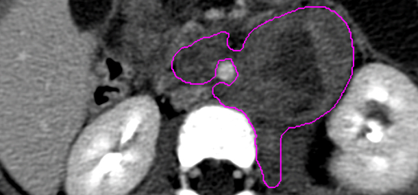
The radiologists at Cincinnati Children’s know that the imaging we do directly impacts your child’s diagnosis, treatment and care plans. Because of this we are continually working to find more effective and better ways for our imaging to positively impact our patients. This year at the RSNA we will present our research that may make a significant difference for children with neuroblastoma. Neuroblastomas are one of the more common tumors diagnosed in children and typically grow somewhere in the chest or abdomen. Neuroblastomas often have an unusual shape because they surround other structures in the body and extend into small spaces. The irregular shape of these tumors can make them hard to measure accurately. Our study looked at the best way to use imaging to measure neuroblastoma and see how the tumor is responding to treatment.

Current guidelines recommend measuring neuroblastomas in three dimensions (top to bottom, side to side and front to back), which is different from other tumors that are usually measured in only one or two dimensions. Even a three-dimensional measurement, however, doesn’t fully capture the commonly irregular shape of neuroblastomas. Our study used special computer software that allowed us to measure actual tumor volume by outlining every extension of the tumor. Changes in this measured tumor volume due to treatment was compared to the changes measured using one dimension, two dimension and three dimension measurements in the same patients. We found that one- and two-dimensional measurements can significantly misrepresent the changes in the tumor size. However, three-dimensional measurements, while not perfect reflections of changes in tumor size, were on average within 7% of the actual change in size of the tumor. Our study suggests that three-dimensional measurements are the best way to measure neuroblastoma tumor changes while receiving treatment.
This project that we will be presenting to the RSNA is just one example of how we are always working to improve the imaging we do so that we can provide your child with the very best care.
Contributed by Dr. Andrew T. Trout and edited by Catherine Leopard (CLS)