Radiology’s Dr. Andrew Trout and former research fellow Vinicius Alves, MD, were featured on RSNA News. The article “US Performs Comparably with MR Imaging for Quantifying Fatty Liver Disease in Children” by Evonne Acevedo showcases the research performed at Cincinnati Children’s explains how “Imaging can be used to rule out other causes of liver disease.”

The article was aimed at radiologists, but here we provide a concise explanation aimed at patients and families to share the importance of the research.
Imaging Fatty Liver Disease in Children
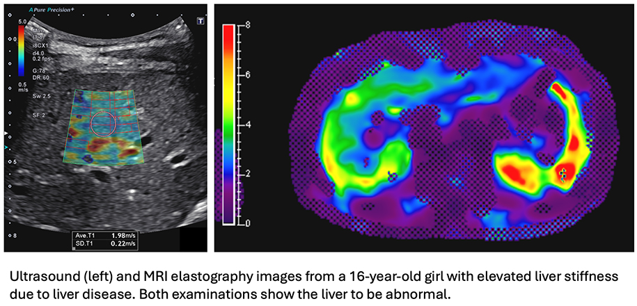
Metabolic dysfunction-associated steatotic liver disease (MASLD)—formerly known as Non-Alcoholic Fatty Liver Disease (NAFLD)—is increasingly prevalent among young patients. In the prospective study by the team at Cincinnati Children’s, researchers explored using both ultrasound (US) and MR imaging to diagnosis MASLD, providing valuable insights for clinicians and families.
Why Is This Important?
Conclusion
Both US and MR imaging are reasonable alternative methods for noninvasively quantifying liver disease in children with a body mass index less than 35 kg/m². Using either technique, clinicians may be able to enhance early detection and management of MASLD, ultimately benefiting our patients.
This study underscores the importance of accessible and accurate imaging tools in safeguarding the health of our young patients. By utilizing both US and MR imaging effectively, we can make informed decisions and improve outcomes for children at risk of fatty liver disease1.