The opportunity to partner with the Cincinnati Museum Center and to be part of history at Cincinnati Children’s last night was an honor and an exciting opportunity as our team of radiologists and technologists performed the virtual autopsy of a Peruvian child, mummified since around the 15th century.
This is the first time I have been involved in the imaging of a child mummy and the first time such imaging has been performed at Cincinnati Children’s. We were approached to take part in this project in conjunction with the museum’s current exhibit, “Mummies of the World,” in order to discover more about this child’s life and ultimate death.
For me and my colleagues in the radiology department, being involved in this project presented an exciting and intellectually stimulating opportunity in line with the scientific missions of both our institutions. In addition, it was an opportunity for us to engage with the community and advance the understanding of the history of this child and its culture of origin.
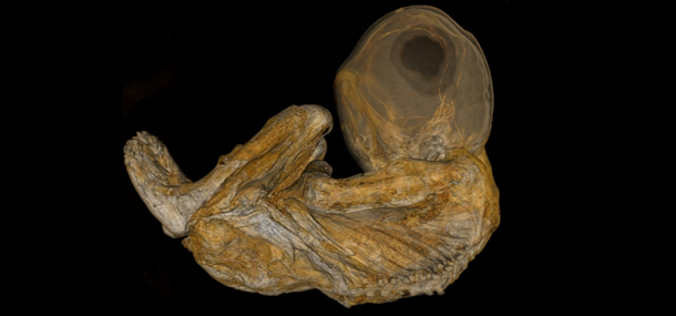
Prior to the examination, we and the museum staff knew only a little about the mummy, who is believed to have been buried 400 to 500 years ago. Unlike some mummies which are deliberately preserved and wrapped, this mummy had been preserved naturally due to the conditions where it was buried. The level of preservation of this mummy is spectacular. While the eyes are no longer present, much of the skin including ears is intact, although dried out.
One particular area of interest about this mummy is the elongated shape of the child’s head. Through the course of our examination, we hope to determine whether this shape was due to intentional modification, in line with cultural norms of the time and region of the world, or if the shape resulted from a developmental abnormality.
Handling the child with respect and care was of utmost importance to our staff, the museum, and the Mummies of the World exhibit curators. Together, we took precautions along every step of the way to protect the mummy and ensure the safety of our patients and families during the course of the examination.
Over the next few weeks, we will be working with the Cincinnati Museum Center and the curators to examine the more than 12,000 images we have generated to learn more about this child. We hope to be able to determine the sex of the child, any ailments or abnormalities the child may have had, and potentially the child’s cause of death.
See the initial images from the scans and learn more about the types of scans we used. And you can also find radiology on Twitter at @CincyKidsRad, where last evening’s events were captured and shared in detail as they happened.
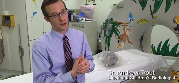
Dr. Andrew Trout is an assistant professor of Radiology and interim director of Thoracoabdominal Imaging. His areas of clinical practice include general pediatric radiology, thoracoabdominal imaging and nuclear medicine.