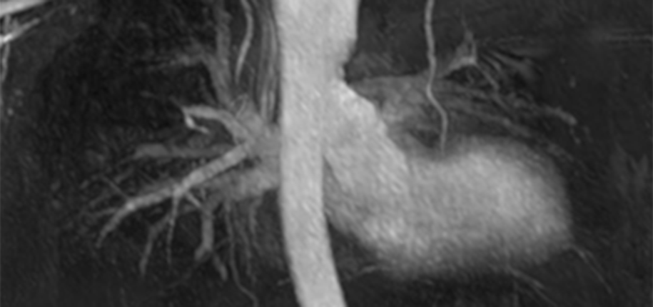
Your body has vessels called arteries and veins that carry blood to and from every part of your body. It takes only about one minute for your blood to travel through the entire body and back to the heart! Sometimes, though, vessels can develop abnormalities such as an aneurysm, clot, narrowing, or other malformation. In the Radiology department, there are many ways we can look at these vessels that carry vital oxygen and nutrients to every organ in your body and help diagnose problems that may arise.
There are several modalities in the Radiology department that can be used to image blood vessels, and our radiologists work with your doctor to determine which test is the best for you or your child. Potential tests used to map the blood vessels include ultrasound, CT, MRI and interventional radiology.
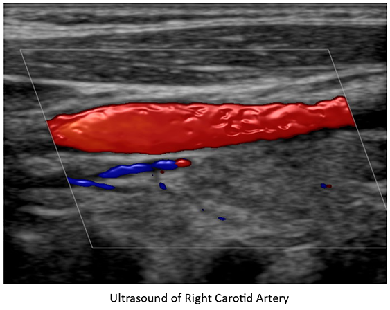
During an ultrasound, the technologist will use warm jelly with a handheld transducer to look through your skin at the vessel. You may be asked to hold your breath or move in different positions during the exam.
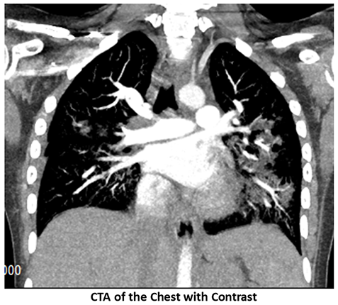
When you have a CT scan, you will be given a contrast injection through an intravenous line (IV), which is placed in your arm by your technologist. The technologist will take your pictures at the exact right time to look at a particular vessel.
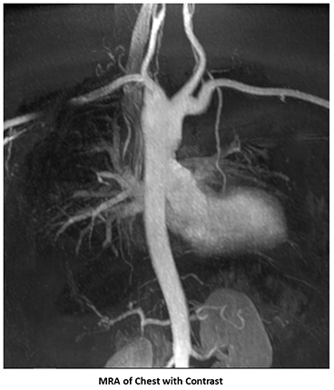
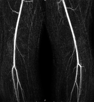
In MRI, there are instances when we use a contrast, or some vessels can be imaged without using any contrast at all. If your doctor is mainly interested in seeing what the vessels look like, we can get a good overall look without using contrast. Special techniques allow the scanner to image only the vessels, and then the MR technologist can manipulate the data to allow the vessel to be viewed from all different angles. Sometimes your doctor wants to see how the vessels work to carry the blood, so you might have a dynamic contrast-enhanced MRA. The technologist will inject a little bit of contrast through an IV in your arm and take timed images of the area, watching the contrast fill the arteries into the area and then go back out through the veins.
In an interventional procedure, the contrast is given through a tiny tube, which is placed by the radiologist straight into the artery or vein itself. We use special cameras to watch the blood flow in motion to look for any areas that may be blocked or where the blood cannot flow properly. In most cases, the doctor can fix problems at that time, using clips or coils, special glue, balloons, or by placing a stent.
If your doctor should ever want to get a good look at your blood vessels, our Radiology team at Cincinnati Children’s Hospital will help determine which test is the best option for you. No matter which area you end up visiting, you can be assured that you will be given the best care by our excellent imaging team!