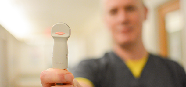
Photo: Ultrasound technologist holding a transducer
Each year, 15 million babies worldwide are born too soon. In the United States, 1 in every 9 babies is born premature. This is significant because premature birth is the number one cause of death in infants and is a leading cause of lasting childhood disabilities.
If a baby is born premature, he or she will be treated and monitored in the neonatal intensive care unit. Due to their immature state, these babies are at risk of many complications, including brain problems. When the doctors are concerned about the baby’s brain, we as radiologists and ultrasound technologists at Cincinnati Children’s can help. At this early age, the brain can be seen with ultrasound through the baby’s soft spot on the top of the head. In addition, the ultrasound equipment is portable, so we can come to the unit and perform the exam at bed side; the exam does not require sedation. One last important advantage of ultrasound is that it can provide answers without any need of radiation, making it the perfect screening tool for these children.
The procedure starts with the ultrasound technologist gently applying a warm gel over the baby’s soft spot to get better contact between the baby’s skin and a hand-held wand (the transducer). She then uses the wand to start collecting the images that will be saved for the radiologist to review. Then, the radiologist reads the images and lets the baby’s doctors know if it is normal or if a complication has been found. Premature babies are at risk of bleeding in the brain, and if this is found on ultrasound, we will be performing close follow ups to monitor resolution or detect potential complications as early as possible so the doctors can act on them.
Contributed by Dr. Maria Calvo-Garcia and edited by Sarah Kaupp (RRA).