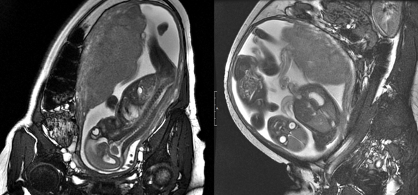
Many people might not know that ultrasound isn’t the only way to image an unborn child. Here at Cincinnati Children’s, we perform MRIs on pregnant women almost every day.
MRI (magnetic resonance imaging) is a way of taking detailed pictures of the inside of the body using a magnetic field combined with pulses of radio wave energy. Like ultrasounds, there is no iodizing radiation emitted from the MRI; therefore, there are no concerns about exposure to the unborn child.
Utilizing the exact same technology used for imaging all other body parts, we are able to perform an MRI to look at the fetus, or unborn child, in the uterus during second- and third-trimester pregnancies. This allows us, the radiologists, to add valuable information to the findings already identified on obstetrical ultrasound. Fetal MRI is of particular value when looking at the brain of the fetus and has allowed us to identify and better characterize abnormalities even before the child’s first breath. Plans and treatments can begin weeks to months before the birthdate, allowing the best care to start as soon as possible.
We are extremely fortunate to work in conjunction with the Cincinnati Fetal Center in collaboration with University of Cincinnati Medical Center and Good Samaritan Hospital, which brings together a network of physicians, nurses and technologists who specialize in diagnosing and treating complex and rare fetal conditions. With over 10 years of experience in fetal MRI, we strive to provide the best images and interpretations possible so that physicians can provide the earliest care and counseling to our patients. With continued advances in imaging and surgical techniques, we hope to improve outcomes in high-risk pregnancies so that every child has a fighting chance of beginning life with the best possibilities.
Contributed by Dr. Usha D. Nagaraj and edited by Tony Dandino (RT).