At a recent research showcase, Cincinnati Children’s Artificial Intelligence Imaging Research Center (CAIIR) highlighted its cutting-edge work in pediatric imaging innovation. Among the presentations was a poster by research assistant Alexandra J. Hendricks-David, detailing a groundbreaking study commissioned by the Center for Pulmonary Imaging Research (CPIR). The focus was to develop an automated segmentation model of the lung and airways to process UTE MRI images, a potential alternative to CT scans for monitoring tracheal collapse in NICU patients.
Why UTE MRI Matters for Neonates
CT imaging, while highly detailed, involves radiation exposure, a concern for neonatal patients due to their small size and vulnerability. UTE (Ultrashort Echo Time) MRI offers a radiation-free alternative, making it a promising tool for routine monitoring of airway conditions like tracheal collapse. Though MRI lacks the fine resolution of CT for pulmonary tissues, it can be used proactively to detect early signs of collapse, reserving CT for more detailed follow-up when necessary.
The Challenge of Manual Segmentation
Each UTE MRI 3D image is comprised of hundreds of 2D slices, which traditionally require manual segmentation of a labor-intensive process where researchers outline organ boundaries slice by slice. This task, while essential, consumes valuable time and expertise that could be better applied elsewhere.
Enter AI: Automating the Segmentation Process
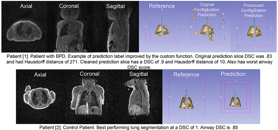
To streamline this process, the IRC leveraged its expertise in AI model development for medical imaging. Using the MONAI Project, an open-source framework for healthcare imaging AI, the team experimented with various prepackaged model architectures to automate segmentation. Their goal: create the first AI model specialized in airway and lung segmentation for NICU patients with diverse pulmonary pathologies. The CPIR study involved UTE MRI scans of 153 NICU patients, producing 413 3D image stacks. This dataset was used to develop and validate a deep-learning-based AI model.
Filling a Critical Research Gap
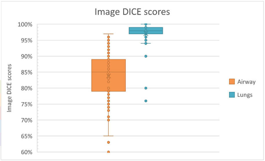
The nnunet-v2 model gave human-level lung segmentations with a similarity comparison of 97% overall. The model did not produce the same quality results with the airway segmentations compared to the lung; this was not a surprise to our researcher since the airway has large inter-reader variability amongst human segmentators as well. The airway images in this project were more challenging than average due to the patient size (being neonates) and pathology.
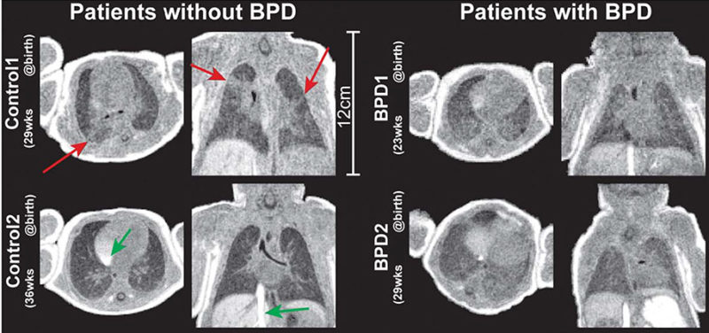
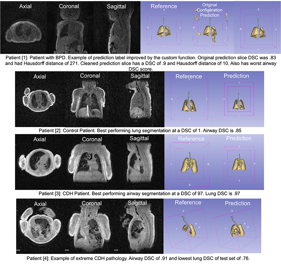
A literature review revealed a striking gap in neonatal-focused segmentation studies. Only two European papers addressed similar challenges; one on bronchopulmonary dysplasia (BPD) and another on in vitro segmentation of fetal lungs and liver with CDH. The IRC’s work stands to be the first published model targeting airway and lung segmentation across a broad NICU population, marking a significant advancement in pediatric imaging research.
Bronchopulmonary Dysplasia (BPD): Also known as respiratory distress syndrome. Usually affects infants born 10+ weeks premature and exhibits breathing issues at birth. Serious BPD is usually caused by underdeveloped pulmonary organs – lungs do not produce a surfactant to keep lungs open
Congenital Diaphragmatic Hernia (CDH): Diaphragm contains opening that allows abdominal organs to displace into chest cavity – limited space reduces space for lung development in vitro. Causes breathing complications
Pulmonary Hypoplasia (PH): Underdeveloped or irregularly developed lungs. Comorbid with other conditions, but some cases do not have obvious underlying cause
Tracheoesophageal Defect (TED): Trachea and esophagus abnormally develop. This grouping includes various tracheal and esophageal defects.
Ali-Joy Hendricks is a research assistant, working in Imaging Research Center of Cincinnati Children's.
Glenn Miñano is a media specialist in the Department of Radiology, providing graphic design, photography, printing, video services, and administration of the department’s online properties. His works have been published in several medical articles, such as the American Journal of Radiology and the American Institute of Ultrasound. He has been providing these services to the Radiology Department since 1996.
Meredith Towbin is a freelance copy editor and writer. She has copyedited the Department of Radiology’s blog since it launched. She also works as a copy editor for the home improvement website BobVila.com. Her writing has been featured on HuffPost as well as other writing sites.