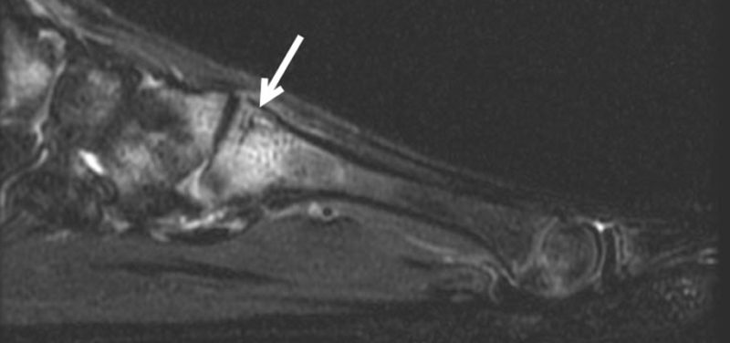

Featured Image: T2 image lateral view shows the bright signal and dark fracture line stress fracture (arrow)
With spring and warmer weather right around the corner, sports seasons change and children’s physical activity levels may increase. Bones constantly repair and rebuild in accordance with the physical stresses placed upon them. Early season training in particular puts new stresses on the developing musculoskeletal system. Even healthy bones may be at risk for fracture when subjected to repetitive stress if it exceeds the bone’s capacity to remodel.

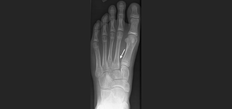
Image: X-ray – no fracture is seen in area of symptoms (arrow)
Complaints of lower extremity or foot pain are common several weeks into training for sports that involve running, jumping, and change of direction (track, basketball, tennis, gymnastics, etc). These symptoms may become more constant with the repetitive activity. Girls are at higher risk than boys, and the site of stress injury will vary somewhat with activity. Stress fractures most frequently occur in the shin bone (tibia) or the bones of the foot but may even occur at the hip (femur). Those in the metatarsal bones of the foot have been called “march fractures”, as they were described in soldiers who sustained the injury after prolonged marching.

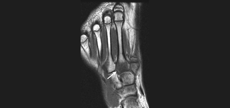
Image: T1 image frontal view shows the darker abnormal signal in the base of the 2nd metatarsal (arrow)
If medical attention is needed, x-rays will often appear normal in the early stages of a stress fracture since only a small portion of the bone may be involved. X-rays performed over 2 weeks after the onset of symptoms are more likely to show signs of a healing response that may confirm the diagnosis. MRI is much more sensitive than x-rays for the detection of stress injuries in bone, and occasionally may be necessary to make the diagnosis in the setting of normal x-rays or concern for other soft tissue injuries that may be present.
The good news is that these fractures will heal with rest. The amount of time to heal is dependent on the location and severity of the injury.
Contributed by Dr. Kathleen Emery and edited by Janet Adams, (ADV TECH-US).
